#Product Trends
Bladder Space-Occupying Lesion
Case Sharing
· Patient information:
62 year male patient complaints of intermittent gross hematuria for 2 weeks and dysuria with urinary retention in the past 2 days.
· Routine urine test:
urinary red blood cell count (RBC) 432.8/ul, urinary red blood cell (RBC) 78.6/HPF.
· History:
10 years of hypertension.
· Ultrasonic findings:
space occupying lesion in bladder.
Difficulties and differential diagnosis of cases
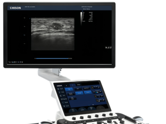
Ultrasonography showed a space-occupying lesion in the bladder. Given the need for clear pathological diagnosis in disease staging. The ultrasound image clearly show the lesions and there is a safe puncture path, and the patient's general condition and test results meets the basic condition for implementation and completion of puncture. Bladder puncture biopsy (18G puncture needle) is used for diagnosis.
Because the bladder can store urine, ultrasound can easily show the protrusion lesions of the bladder wall and the objects in the bladder, so it can be used as an important method to survey bladder diseases. However, it is difficult to determine the nature of the solid small protrusion lesions by other ultrasonic examinations. If the mass is small and the bladder wall where the mass is located is smooth, it is difficult to determine the nature of ultrasound when the bladder wall is invaded by malignant tumors at the early stage. Therefore, cystoscopy and pathology should be combined to make a clear diagnosis when ultrasound finds space-occupying lesions, to avoid misdiagnosis.
Ultrasonographic manifestations of normal bladder
When the bladder is full, the urine in the bladder is an echo-free area, the bladder wall is a bright echo light band, the mucosal inner wall and the urine interface reflect strong echo, which is a flat thin light band, and the submucosal muscle layer is uniform, medium, and low echo.
Diagnostic value of ultrasonography in bladder space-occupying lesions
Bladder space-occupying lesion is one of the common diseases of the urinary system. Correct diagnosis is of great significance to guide clinical treatment. Ultrasonography can determine the shape, scope, and internal echo of bladder lesions, judge whether the lesions have infiltrated the bladder wall and muscle layer, and the extent of infiltration according to whether the bladder contour of the attachment site of the lesions is complete or not, and also judge whether there are adjacent organs and lymph node metastasis, so it is still the preferred method for screening bladder space-occupying lesions. Ultrasonography is of great clinical significance in formulating appropriate treatment plans, in preoperative preparation and intraoperative management, and the diagnosis of prognosis.
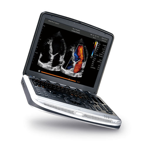
Medical imaging technology has developed rapidly. Because of its small size and portability, portable ultrasound can be scanned in any scene, making it gradually applicable in various clinical departments. The SonoBook series of portable color ultrasounds is as small and light as a laptop and has high-definition image processing technology. At the same time, it has varied probe configurations to help doctors scan and diagnose bladder space-occupying lesions more accurately.