#Industry News
Interview to Dr. Grant Myhre: "Why did you decide to have an MRI?"
Why is MRI technology so crucial for detecting specific pathologies?
Why is MRI technology so crucial for detecting specific pathologies?
“Seeing” the inside of a horse’s foot can be a struggle. Initially, we used radiographs (x-rays), but the images did not show enough soft tissue detail. We moved on to thermography which looks at the heat in the hoof but is not particularly useful otherwise. Using ultrasound through the hoof was an improvement but does not compare to the clarity provided by MRI.
The gold standard for lameness detection
Ninety percent of lameness in the front leg is due to pathology in the foot. One of the most challenging diagnoses in a horse is injury to the deep digital flexor tendon. You cannot visualize this well without an MRI. MRI can give a diagnosis, determine prognosis, and help guide treatment of the injury.
A neurectomy can be an excellent treatment option. Using the MRI, we can determine the extent of degeneration of an injury. The extent of injury determines how much exercise the horse can return to after treatment and gives a long-term prognosis of the injury.
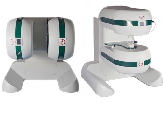
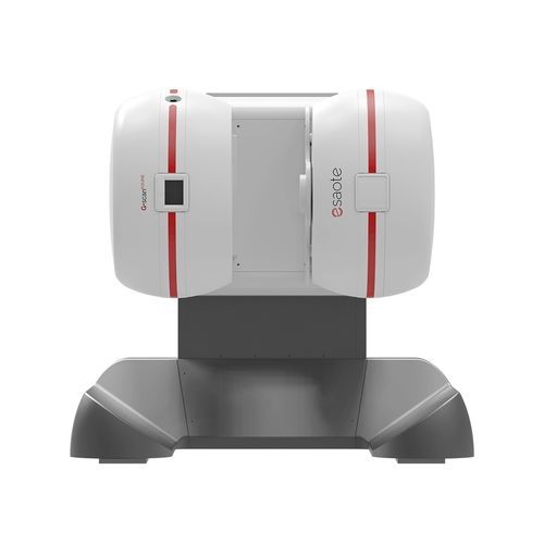
Why did you choose the G-scan equine for your clinic?
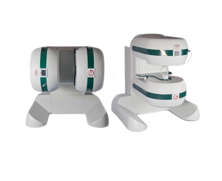
Before we installed the G-scan equine, I used the ELLEGRO system from Esaote for many years to scan equine limbs. It was great for our needs at the time, and due to our experience working with the company and the versatility and flexibility of their systems, it was easy to upgrade to the G-scan equine for the improvements in image quality. When we scan limbs under general anesthesia, we can acquire crisp, clinical images. If the horse is not a candidate for anesthesia or the owner is opposed, we can rotate the magnet 90 degrees which allows for a standing exam.
We also have specialized coils to help visualize what’s wrong, ranging from areas of interest in the hoof all the way to the head. One of my favorite features about this system is the ability to image the stifle, which most other scanners cannot do!
How do you perform stifle examinations?
The rotating G-Scan equine is turned 90 degrees into a vertical position. We put the horse in dorsal recumbency (on his/her back) on the MRI table. The affected leg is then anchored in the magnet’s isocenter, with the other leg straddling the MRI.
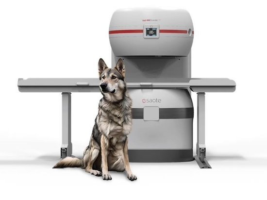
How was the G-scan equine installation experience for your clinic?
Since we previously installed the ELLEGRO system, I was familiar with the ease of the installation process. The G-scan equine size conveniently fits within our space requirements and our business strategy. High-field MRI requires significant expense for construction, helium, energy, and maintenance costs. This dedicated MRI from Esaote is safe, has a smaller footprint, and allows us to lower costs for our clients.
For horses, I have everything I need.