#Industry News
DDR - Dynamic Digital Radiography
Dynamic Digital Radiography (DDR) is a novel X-ray technology that provides a series of individual digital images acquired at high speed and low dose.
The resulting cine loop enables clinicians to observe the dynamic motion of anatomical structures over time, enhancing diagnostic capabilities.
DDR is the only imaging technology that provides a view of anatomy in motion, with a large field of view and low radiation dose.
DDR images can be analyzed and quantified with advanced image processing of the DI-X1 workstation providing additional functional information of anatomical structures and therefore increasing the diagnostic capabilities in several clinical areas.
Therapy assessment of respiratory diseases:
DDR provides quantitative information related to the lung area and the diaphragm excursion that can be used for the therapy evaluation of several respiratory diseases like COPD, asthma, etc.
Tracheal and Diaphragm motion evaluation:
Moreover, we can identify disfunctions related to the physiological movement of the anatomical structure, like in case of diaphragm palsy or tracheal narrowing
Optimize Surgical Planning:
DDR can provide Respiratory Motion Visualization useful for assessing adhesions and invasions, based on lack of lung tissue motion, difficult to observe with CT or other modalities. Determining the existence of adhesions makes possible the selection of best surgical procedure such as thoracotomy or thoracoscopic surgery.
Post-Operative Evaluation:
DDR can be a potential technique for post-operative evaluation to assess and follow-up how organs are recovering after a lung resection, for example.
Detection of Perfusion Defects
DDR exhibits its potential in the cardiovascular field as well because it allows to obtain functional information of lungs, based on signal changes related to pulmonary circulation and ventilation without use of IV contrast or radiopharmaceutical. Thus, DDR might be a diagnostic tool to identify perfusions defects correlated to pulmonary thromboembolism.
Detection of Perfusion Defects
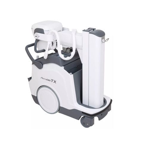
Mobile Aero DR TX will enable DDR capabilities, brining a huge potential use of DDR for patient management and follow-up in ICU departments where it can be used for the assessment of ventilation, detecting perfusion defects related to pulmonary thromboembolism and more evaluations that can be done without patient transferring!
Motion assessment:
DDR is useful for assessment of joint motion and for spine stability. It is possible to perform acquisition with joints doing movements repeated during daily routine and can be useful for treatment follow-up and pos-operative evaluation.
DDR main benefits
Clinical value – DDR presents a 17" x 17" view of quantifiable and functional findings not detected with standard X-ray
Accessibility– DDR capabilities with the new mobile AeroDR Tx enables portable exam anytime and anywhere in ICU department and/or patient ward improving diagnostic capabilities
Low cost – DDR may reduce the need for advanced imaging like CT, MRI and scintigraphy.
Patient-centric – DDR uses low radiation, does not require contrast, and can be performed in multiple positions
Efficient – Full exams are performed in minutes and performed as standard x-ray
Effective – Visualize and quantify movement, interaction, dynamic density, change over time, and pathophysiology
Boost your clinical practice to the next level