#Industry News
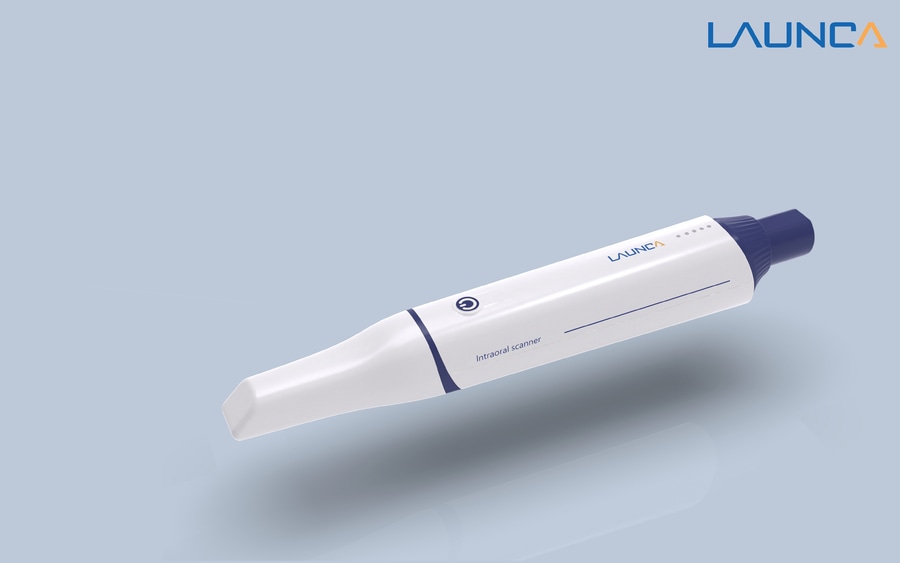
How to Use the Launca DL-300 Wireless to Scan the Last Molar
Step-by-Step Guide to Scanning the Last Molar
Step 1: Prepare the Patient
Positioning: Ensure the patient is comfortably seated in the dental chair with their head properly supported. The patient's mouth should be opened wide enough to provide clear access to the last molar.
Lighting: Good lighting is crucial for an accurate scan. Adjust the dental chair light to ensure it illuminates the area around the last molar.
Drying the Area: Excess saliva can interfere with the scanning process. Use a dental air syringe or a saliva ejector to keep the area around the last molar dry.
Step 2: Prepare the Launca DL-300 Wireless Scanner
Check the Scanner: Ensure the Launca DL-300 Wireless is fully charged and the scanner head is clean. A dirty scanner can result in poor image quality.
Software Setup: Open the scanning software on your computer or tablet. Make sure the Launca DL-300 Wireless is properly connected and recognized by the software.
Step 3: Start the Scanning Process
Position the Scanner: Begin by positioning the scanner in the patient's mouth, starting from the second-to-last molar and moving towards the last molar. This approach helps in getting a broader view and a smooth transition to the last molar.
Angle and Distance: Hold the scanner at an appropriate angle to capture the occlusal surface of the last molar. Maintain a consistent distance from the tooth to avoid blurry images.
Steady Movement: Move the scanner slowly and steadily. Avoid abrupt movements, as they can distort the scan. Ensure you capture all surfaces of the last molar – occlusal, buccal, and lingual.
Step 4: Capture Multiple Angles
Buccal Surface: Start by scanning the buccal surface of the last molar. Angle the scanner to ensure the entire surface is captured, moving it from the gingival margin to the occlusal surface.
Occlusal Surface: Next, move the scanner to capture the occlusal surface. Ensure the scanner head covers the entire chewing surface, including the grooves and cusps.
Lingual Surface: Finally, position the scanner to capture the lingual surface. This might require adjusting the patient's head slightly or using a cheek retractor for better access.
Step 5: Review the Scan
Check for Completeness: Review the scan on the software to ensure all surfaces of the last molar are captured. Look for any missing areas or distortions.
Rescan if Necessary: If any part of the scan is incomplete or unclear, reposition the scanner and capture the missing details. The software often allows you to add to an existing scan without starting over.
Step 6: Save and Process the Scan
Save the Scan: Once satisfied with the scan, save the file using a clear and descriptive name for easy identification.
Post-Processing: Use the software's post-processing features to enhance the scan. This might include adjusting brightness, contrast, or filling in minor gaps.


