#Industry News
Correct Lead Placement in ECG: A Complete Guide for Accurate Readings
woman 12 lead ecg placement female
Introduction to ECG and Its Importance
Electrocardiography (ECG or EKG) is one of the most vital diagnostic tools in modern medicine. By recording the electrical activity of the heart, it allows doctors to detect arrhythmias, myocardial infarctions, ischemia, and many other cardiac conditions. However, the accuracy of an ECG heavily depends on correct lead placement. Even a small error can distort the tracing, leading to misdiagnosis and inappropriate treatment.
This guide explains everything you need to know about lead placement in ECG, from the basics of the 12-lead system to advanced techniques for special cases.
Understanding ECG Leads
Limb Leads (I, II, III, aVR, aVL, aVF)
Limb leads measure the electrical activity of the heart in the frontal plane.
Lead I: right arm to left arm
Lead II: right arm to left leg
Lead III: left arm to left leg
Augmented leads (aVR, aVL, aVF): measure activity from one limb relative to the average of the others
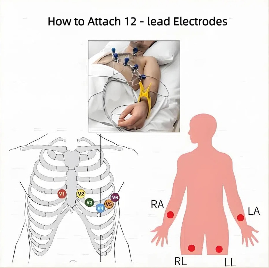
Chest (Precordial) Leads (V1–V6)
Chest leads examine the electrical activity in the horizontal plane. They provide detailed information about the anterior, lateral, and septal walls of the heart.
Standard 12-Lead ECG System
The Difference Between Leads and Electrodes
Many people confuse the terms lead and electrode. Electrodes are the sticky patches placed on the skin, while leads are the electrical views of the heart generated by these electrodes.
Clinical Relevance of the 12-Lead ECG
The 12-lead ECG provides a 3D view of the heart’s activity, making it possible to detect ischemia, infarctions, arrhythmias, and structural abnormalities.
Step-by-Step Guide to Correct Lead Placement in ECG
Preparation Before Placing Leads
Ensure the skin is clean and dry
Shave excessive hair if necessary
Place the patient in a comfortable, supine position
Limb Lead Placement Explained
Right Arm (RA): on the right wrist or shoulder
Left Arm (LA): on the left wrist or shoulder
Right Leg (RL): used as a ground electrode, placed on the ankle
Left Leg (LL): on the left ankle or thigh
Chest Lead Placement (V1–V6)
V1: 4th intercostal space, right sternal border
V2: 4th intercostal space, left sternal border
V3: Midway between V2 and V4
V4: 5th intercostal space, midclavicular line
V5: Horizontal with V4, anterior axillary line
V6: Horizontal with V4, midaxillary line
Common Mistakes in Lead Placement and How to Avoid Them
Swapping limb leads (RA with LA)
Incorrect intercostal space identification
Misalignment of chest leads, especially in obese patients
Placing V1 and V2 too high, which alters ST-segment readings
The Impact of Incorrect Lead Placement on ECG Interpretation
Wrong placement can lead to false diagnosis of conditions like:
Myocardial infarction
Bundle branch blocks
Chamber enlargement
Misinterpretation of axis deviation
Special Considerations in Lead Placement
ECG Placement in Obese Patients
Extra tissue may make anatomical landmarks harder to locate. Leads may need slight adjustments for accurate results.
ECG Placement in Women
Electrodes should be placed beneath the breast tissue when possible to avoid displacement.
Pediatric Lead Placement
Children often require modified lead positions due to their smaller chest size.
Advanced Lead Placements Beyond Standard ECG
Right-Sided Chest Leads
Used when right ventricular infarction is suspected. Leads V3R–V6R are positioned on the right side of the chest.
Posterior Leads
Applied to detect posterior wall infarction. Leads V7–V9 are placed on the patient’s back.
Tips for Healthcare Professionals to Master ECG Lead Placement
Practice anatomical landmark identification
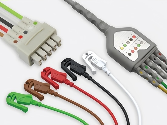
Use color-coded lead wires if available
Confirm electrode adhesion for signal quality
Double-check positions before recording
FAQs on Lead Placement in ECG
Q1. Why is correct lead placement in ECG so important?
Because even a slight error can lead to misdiagnosis of serious cardiac conditions.
Q2. Can ECG be done with fewer than 12 leads?
Yes, but the 12-lead ECG provides the most complete diagnostic view.
Q3. What happens if V1 and V2 are placed too high?
It can mimic right bundle branch block or anterior wall ischemia.
Q4. How do I place ECG leads on a female patient with large breasts?
Move electrodes slightly under the breast tissue, ensuring proper intercostal space alignment.
Q5. Is it okay to place limb leads on the torso?
Yes, in emergencies, limb leads can be placed proximally on the torso, though interpretation may vary slightly.
Q6. Do children need different ECG lead placements?
Yes, children often require adjusted electrode placement due to their smaller anatomy.
Conclusion
Correct lead placement in ECG is a fundamental skill every healthcare provider must master. It ensures accurate readings, avoids misdiagnosis, and ultimately improves patient outcomes. Whether dealing with standard 12-lead setups, special considerations, or advanced placements, attention to detail is critical.