#Product Trends
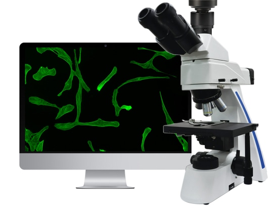
Live cell imaging under Inverted microscope
Phase contrast, fluorescence
In routine laboratory work, culture living cells are available observed under phase contrast microscope, and directly record dynamic changing of live cells by microscope camera with time-lapse captured images. It is more obviously to be seen after staining. Stain live cell with fluorochorme, it can show out beautiful and bright fluorescence excited by fluorescence illumination.
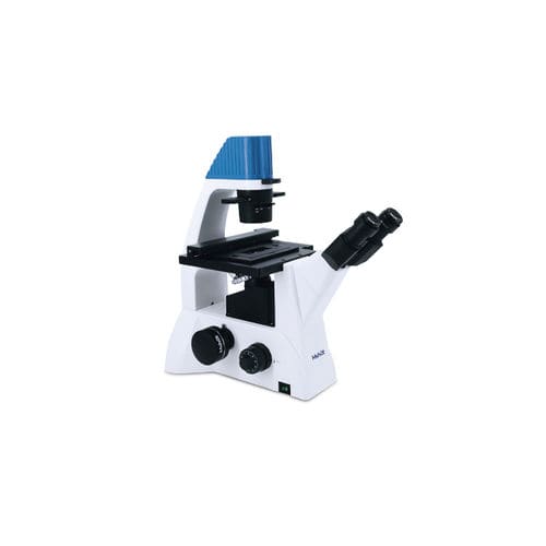
Living cells is transparent when light pass through, the wavelength and amplitude almost no changes. So basic light microscope can not see the living cells without staining. In order to observe the structure of living cells, it is needed to improve the contrast of the structure through other ways. In 1930s, the Holland physicist Zernike design of the phase contrast microscope using light diffraction and disturbing. With more requirements of experiment, people expanded simple phase contrast microscope with fluorescence illumination. Those microscope is called inverted fluorescence microscope.
Professor at Jiangnan University cultivates living cells, using MSHOT Inverted fluorescence microscope MF52-LED and scientific camera MS23 at their labs, to record live cells changing and doing analysis, measure cell sizes.