#Product Trends
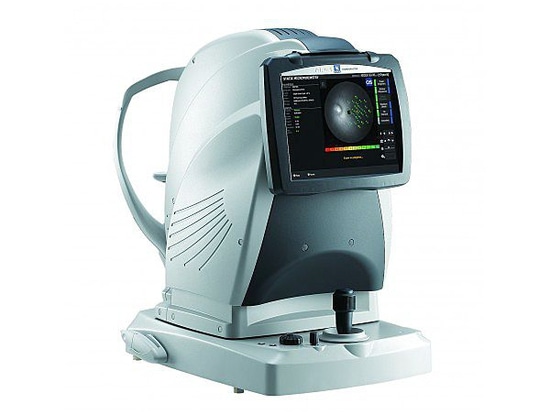
Retina Scan Duo™ by NIDEK - 3D auto tracking, auto shot and user friendly interface
Easy operation with 3-D auto tracking, auto shot, and user friendly interface
High definition images
Wide area scan (12 x 9 mm) / Wide area normative database (9 x 9 mm)
Multiple OCT scan patterns
Value added features
Various reports
The acclaimed 3-D auto tracking and auto shot functions allow easy imaging of the fundus and all its features. Each standard and professional mode has a different image capture interface which can be selected based on clinic preference.
High definition images :
For OCT imaging, up to 50 images can be averaged and the OCT sensitivity is selectable among ultra fine, fine, and regular sensitivities based on ocular pathology. The Retina Scan Duo™ has a built-in 12-megapixel CCD camera, producing high quality fundus images.
Wide area scan (12 x 9 mm) / Wide area normative database (9 x 9 mm) :
A 12 x 9 mm wide area image centered on the macula can be captured with the Retina Scan Duo™. The 9 x 9 mm normative database provides a color-coded map indicating distribution range of the patient''s macular thickness in a population of normal eyes.
Multiple OCT scan patterns :
A wide range of scanning patterns are available to allow the practitioner to select a scan that suits the retinal region and ocular pathology.
Value added features :
Additional value added features include fundus autofluorescence (FAF) and En face OCT.