#Product Trends
Unexplained Dyspnea and Exercise Intolerance: What Can Exercise Hemodynamics Reveal?
Understanding the cardiovascular mechanisms that may remain hidden during conventional exercise testing
Dyspnea and exercise intolerance are among the most common reasons for referral to cardiology, pulmonology, and exercise testing laboratories. Yet many patients continue to experience significant symptoms despite apparently normal resting investigations. Echocardiography, electrocardiography, pulmonary function testing, and even conventional exercise testing may fail to fully explain why a patient remains limited during physical activity.
This discrepancy between symptoms and objective findings represents a frequent clinical challenge. Patients may report breathlessness, early fatigue, reduced exercise capacity, or inability to perform daily activities, while standard examinations provide only partial answers. In many cases, the missing information lies not at rest, but in the body's cardiovascular response to exercise.
Exercise capacity depends on the ability of the cardiovascular system to increase oxygen delivery to the working muscles. According to the Fick principle, oxygen uptake is determined by cardiac output and peripheral oxygen extraction. While cardiopulmonary exercise testing (CPET) provides valuable information regarding global exercise performance, it does not directly identify the central hemodynamic mechanisms responsible for exercise limitation.
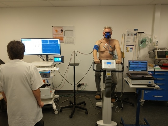
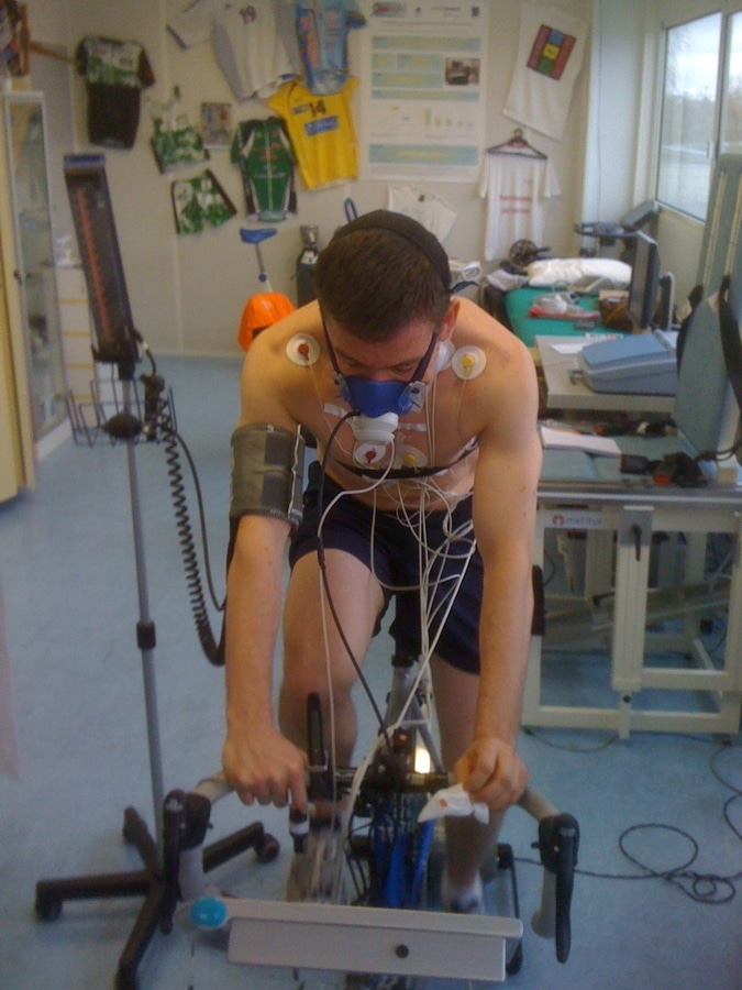
Exercise hemodynamic assessment provides a complementary perspective by continuously monitoring key cardiovascular parameters such as heart rate, stroke volume, cardiac output, cardiac index, ventricular performance, and vascular resistance throughout exercise. These measurements help clinicians understand how the cardiovascular system adapts to increasing workload and whether an adequate hemodynamic reserve is available.
Several distinct physiological profiles may lead to similar symptoms.
Some patients demonstrate an impaired ability to increase stroke volume during exercise, resulting in insufficient augmentation of cardiac output. Others exhibit chronotropic incompetence, where heart rate fails to rise appropriately despite increasing effort. In patients with heart failure, exercise limitation may be associated with reduced contractile reserve and impaired ventricular performance. In contrast, some individuals present with preserved cardiac responses but peripheral limitations related to muscular deconditioning or altered oxygen extraction.
Because these mechanisms may produce similar clinical presentations, understanding the underlying hemodynamic response becomes essential for individualized patient management.
The concept of cardiac reserve is particularly important. Cardiac reserve describes the capacity of the cardiovascular system to increase performance from rest to peak exercise. A patient may present with acceptable resting cardiac function while exhibiting marked limitations in cardiac reserve during effort. Such abnormalities often remain undetected during resting examinations but become evident when cardiovascular responses are assessed dynamically.
A substantial body of scientific literature, encompassing hundreds of peer-reviewed publications, supports the role of exercise hemodynamic assessment as an important component in the physiological evaluation of exercise limitation across a wide range of clinical conditions. Heart failure, unexplained exertional dyspnea, exercise intolerance, pulmonary vascular disease, hypertension, cardiovascular rehabilitation, and sports cardiology are among the areas where exercise hemodynamics has contributed to a better understanding of the mechanisms limiting functional performance.
Contemporary recommendations from the European Association for Cardiovascular Prevention and Rehabilitation (EACPR) and the American Heart Association (AHA) also recognize the importance of cardiovascular and hemodynamic responses during exercise assessment. These recommendations highlight the complementary value of non-invasive cardiac output determination during cardiopulmonary exercise testing and acknowledge the growing clinical and prognostic relevance of bioelectrical methods for assessing cardiovascular function during exercise.
For more than 35 years, PhysioFlow® has contributed to the development and clinical application of exercise hemodynamic assessment. Through collaborations with clinicians, researchers, rehabilitation specialists, and exercise physiologists worldwide, the technology has been used in more than 350 peer-reviewed scientific publications spanning cardiology, pulmonology, exercise physiology, rehabilitation, intensive care, and sports medicine.
Beyond technology itself, the objective remains unchanged: helping clinicians better understand how the cardiovascular system responds to exercise, identify the mechanisms underlying symptoms, and support more individualized patient evaluation and management.
As interest continues to grow in personalized assessment of cardiovascular reserve and functional capacity, exercise hemodynamics provides a unique physiological perspective for understanding why patients become symptomatic during effort, even when conventional resting investigations appear reassuring.