#Product Trends
Detailed Introduction of RFLSI ZW Laser Speckle Contrast Imaging System
An even better tool for microcirculation research.
RFLSI-ZW laser speckle imaging system is an even better tool for microcirculation research based on laser speckle contrast imaging technology (LSCI).
With the advanced optical design and improved image processing algorithm, RFLSI-ZW shows greater performance in imaging field size, image quality, full-field frame rate and optical resolution, and provides a powerful and efficient means for human and animal tissue microcirculation measurement.
Overview:
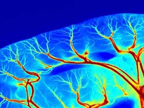
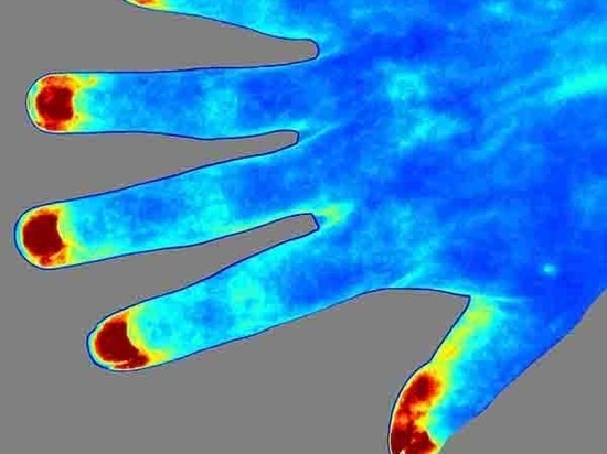
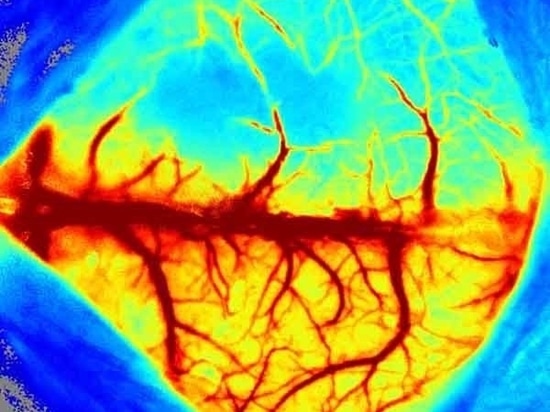
The LSCI technology advantages are its non-contact, high frame-rate, high spatial resolution. They can be used to observe and record blood perfusion of any exposed tissues or organs for microcirculation study or pre-clinical researches like ischemic stroke, lower limbs, mesentery, etc. Multi-Output includes blood perfusion images and videos (500+ million pixels), quantified data for perfusion unit and vessel diameter.
Since 2019, our imaging system has been adopted by more than 100 colleges, universities, and research institutes worldwide such as Stanford University School of Medicine, university of manchester, UC Davis, Duke university. What’s more, it has contributed to publishing more than 60 reputed research papers in magazines like Nature communications, Blood, Diabetes, and Theranostic.
Applications:
Cerebral blood perfusion monitoring
MCAO model assessment
Cortical spreading depression observation
Hind-limb ischemia research
Skin burn/skin flap transplantation
Organ microcirculation observation
Skin allergies
Septic Shock
Chicken Chorioallantoic Membrane Assay
Diabetic Foot
Highlights Of RFLSI-ZW:
1.Image any exposed tissue (skin or surgically exposed tissues) and species.
2.Non-contact, non-contrast agent depending measurement.
3.The built-in CMOS global shutter camera can achieve faster data acquisition and processing speed.
4.Best optical resolution of 3.9 μm/pixel, providing more detailed tissue structures.
5.Max frame rate (full field) up to 100 fps, acquiring real-time changes in larger areas.
6.Motorised 10x optical zoom and auto focus. Image size ranges from 0.57×0.75 to 22.5×30 cm2 in all-in-one imager, covering multiple research applications.
7.Fast auto and fine manual focus, improving focus efficiency and accuracy on various tissues.
8.Optimal lens assembly, filtering the ambient and reflecting light.
9.Class 1 of measurement and indicating lasers, safe to use without eye protection System
10.Laser stability hardware for the ultimate in reliable and consistent measurement over minutes, hours and days.
11.Calibration with calibration box. Self-calibration is possible at any time to keep the equipment in optimal working condition.
12.Trigger In/Out BNC connections for communication with external devices.
13.Unlimited installation of analysis software in PC.