
#Product Trends
Perquisite Chest X-ray Stand for Screening Center How Much Convenience It Brings
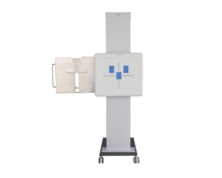
Perquisite Chest X-ray Stand
Chest DR radiography is the most widely adopted and high-value screening procedure among all imaging examinations. Many medical institutions often face common troubles: long queues during peak hours, difficult positioning for patients of varying heights, sluggish mechanical adjustment, ghosting artifacts and high retake rates. Most buyers only focus on the X-ray generator, ignoring the chest X-ray stand as an essential accessory. A reliable chest stand tailored for screening scenarios can directly boost imaging efficiency, reduce retake frequency and improve patient experience. It serves as a hidden workhorse to upgrade the performance of radiology departments.
## 01 Fits Patients of All Ages Enables Fast and Precise Positioning
Screening visitors include the elderly, adults, students and children with a wide range of heights and body types. Ordinary chest stands have limited adjustable travel and cumbersome positioning. For patients with extreme heights, staff have to use footstools or reposition the whole unit, wasting valuable time.
Our chest X-ray stand supports smooth long-stroke vertical adjustment with free fine-tuning. No extra tools are required to complete standing imaging for all crowds. Simple and standardized positioning greatly cuts down the time per examination. It is ideal for high-volume tasks such as corporate group screening, community campaigns and routine check-ups.
## 02 Stable and Clear Imaging Significantly Reduces Retake Rate
Blurriness, ghosting and misalignment are major headaches in screening work. Retakes increase radiation exposure for patients, slow down workflow and cause long waiting queues.
This equipment adopts reinforced integrated frame with solid construction to eliminate shaking during exposure, preventing image distortion and blurred edges. Equipped with professional grid assembly, it effectively filters scattered radiation. Lung markings and thoracic structures are displayed with complete fine details, meeting the requirements for screening and preliminary clinical diagnosis, and lowering retake rates fundamentally.
## 03 Compatible with Mainstream Equipment with Excellent Versatility
Many screening centers mix old and new devices. Mismatch between chest stands, detectors and cassettes is a frequent problem, requiring frequent part replacement and disrupting workflow.
Our chest stand features strong compatibility. It seamlessly works with wired or wireless DR flat panels, CR cassettes and traditional film media. It supports detectors of all common sizes and fits most X-ray and DR units available on the market. One unit supports imaging of chest and thoracic spine, eliminating redundant procurement and cutting capital and maintenance costs.
## 04 Simple Operation Handles Heavy Screening Peak Load
High patient flow and tight schedules are typical during screening peaks. Clunky controls and stuck adjustment mechanisms greatly limit department throughput.
Our chest X-ray stand is designed for simple one-touch adjustment. Lifting and detector loading are straightforward. Radiographers can master operation quickly without lengthy training. Smooth adjustment accelerates patient intake to handle heavy screening workload, shorten waiting time and improve overall service reputation.
## 05 Compliant Radiation Protection Provides Dual Safety for Staff and Patients
Safety is the top priority for medical imaging equipment. Manufactured in line with medical radiation protection standards, the unit is fitted with lead protective curtains to block scattered X-rays and minimize radiation risk for both operators and patients.
The anti-tip stable frame eliminates potential safety hazards. The waterproof and corrosion-resistant surface is easy to clean for hygienic medical environments. It delivers long service life and low failure rate, maintaining stable performance under continuous heavy use for long-term routine operation of screening facilities.
Imaging quality and workflow efficiency do not rely solely on the main X-ray unit. A well-matched professional chest stand is a key detail to raise the standard of radiology departments.
With fast positioning, high-definition imaging, broad compatibility and flexible functions, this screening-specified chest X-ray stand is suitable for imaging centers, clinics, primary hospitals and mobile screening projects. It is a cost-effective solution for medical facilities to optimize service quality and improve operational efficiency.
如果你需要,我可以顺带生成对应的 meta title 和 meta description 用于网页 SEO。
今天 09:36
What Is The Best Handheld X-Ray Machine for Veterinary Use
根据这个题目,围绕 SEO 优化需求,以通俗专业的口吻详解工作原理,同时融入核心知识点、行业常识与实操价值,结构清晰、适配爬虫收录,贴合行业网站发文场景,分段明了,可以有小段落小标题,写一篇字数大约为 1000 字的文章
# What Is The Best Handheld X-Ray Machine for Veterinary Use
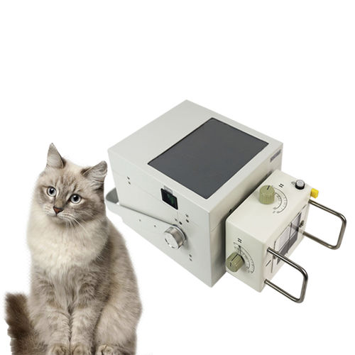
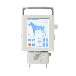
Veterinary clinics and mobile animal care services constantly seek reliable imaging tools to diagnose fractures, internal injuries, and organ abnormalities for companion animals and large livestock. Many practitioners wonder what makes the best handheld X-ray machine for veterinary applications. Unlike fixed stationary X-ray systems, portable handheld veterinary X-ray units are engineered for on-site, point-of-care imaging. The ideal model balances radiation safety, image clarity, lightweight design, and flexible operation for different animal species.
## How a Handheld Veterinary X-Ray Machine Works
A handheld veterinary X-ray generator relies on a compact high-frequency X-ray tube. When powered, electricity accelerates electrons inside the tube, and the collision of electrons with the target produces X-ray photons. These X-rays penetrate the animal’s body. Denser tissues like bones absorb more rays, while soft tissues allow more radiation to pass through. The flat-panel detector captures the remnant X-ray signal and converts it into a digital radiographic image instantly.
The whole system includes three core components: handheld X-ray tube head, digital flat panel detector, and image processing workstation. Operators control exposure through a hand switch. Compared with fixed X-ray equipment, handheld models remove the restriction of dedicated radiographic rooms. This core feature makes it perfect for farm visits, emergency animal rescue, and in-clinic bedside examination.
## Key Features to Judge the Best Handheld Veterinary X-Ray Unit
### 1. Adjustable kV and mA parameters for multi-species imaging
Animals vary greatly in size, from cats and small dogs to horses and cattle. The best handheld veterinary X-ray machine supports adjustable kVp and mA settings. Lower energy parameters work for small companion pets, while higher output penetrates thicker body tissues of large animals. Adjustable output prevents overexposure or underexposure, reducing the rate of retakes and unnecessary radiation exposure for animals and operators.
### 2. Lightweight body with strict radiation shielding
Weight is one of the most practical indicators for handheld devices. Practitioners need to hold the unit steadily during exposure. Excessively heavy equipment leads to hand tremors and blurred images. Meanwhile, built-in lead shielding inside the tube head is non-negotiable. Quality handheld veterinary X-ray machines contain integrated shielding to cut down leakage radiation, protecting veterinarians and assistants during mobile field work.
### 3. Digital DR compatibility and fast image preview
The top-tier handheld X-ray system works seamlessly with wireless flat-panel detectors. After exposure, the radiographic image transfers to the laptop or tablet in seconds. Vets can view the result immediately on site without waiting for film development. DICOM format support allows easy image storage, archiving and remote consultation, which is very helpful for multi-clinic veterinary groups.
## Main Application Scenarios for Handheld Veterinary X-Ray Machines
Mobile veterinary services benefit most from handheld X-ray devices. For equine practices, it is difficult and stressful to transport large horses to a fixed radiography room. A handheld X-ray unit lets veterinarians complete limb radiography directly on the farm. For small animal clinics, it is ideal for emergency cases: checking fractures after trauma, diagnosing bladder stones, or evaluating chest conditions.
It also works for animal shelters and wildlife rescue organizations. When injured wild animals cannot be moved, on-site X-ray imaging helps make rapid diagnosis without aggravating their injuries.
## Practical Tips for Selecting and Operating
When selecting a handheld veterinary X-ray machine, do not only focus on low price. Buyers should verify certification, radiation leakage test reports, and after-sales support. Spare parts availability and on-site training are important for long-term use.
During operation, users must follow local radiation safety regulations. Keep proper distance and use protective aprons, thyroid collars and gloves. Never hold the animal manually during exposure; use positioning aids to avoid occupational radiation exposure. Regular equipment calibration ensures stable image quality and extends the service life of the machine.
## Final Thoughts
The best handheld X-ray machine for veterinary use is not simply the highest-power or cheapest model. It is the equipment that matches your main animal types, workflow and safety requirements. Good handheld veterinary X-ray equipment brings diagnostic convenience, improves the speed of treatment, and reduces stress for animals. For mobile vets and small animal clinics, it is a valuable investment to upgrade diagnostic capabilities.