#Industry News
Atrial Septal Defect: Understanding a Common Heart Condition
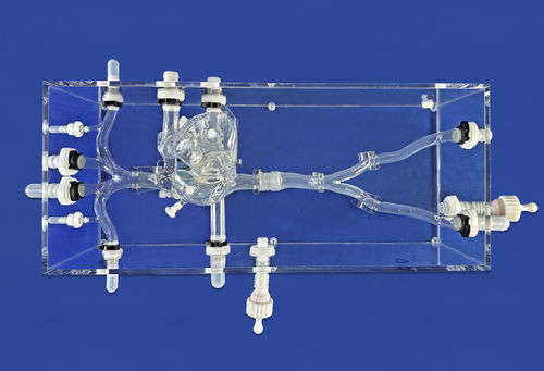
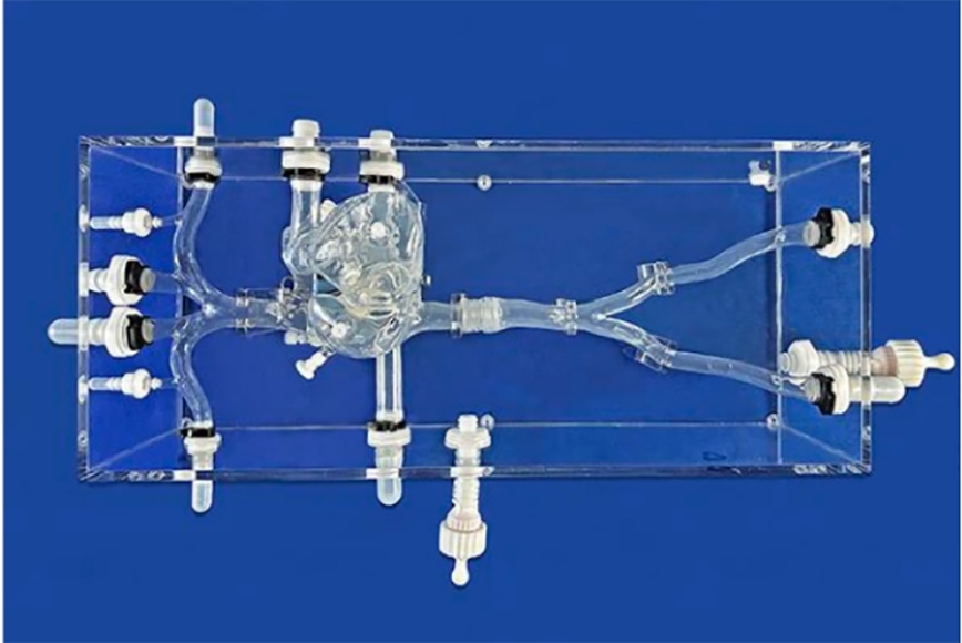
Cardiac Vein With ASD II
An atrial septal defect is a birth defect of the heart in which there is a hole in the wall (septum) that divides the upper chambers (atria) of the heart. A hole can vary in size and may close on its own or may require surgery.
Causes and Types of ASD:
Atrial septal defects most commonly occur due to a combination of genetic and environmental factors. The exact cause is often unknown, but it is believed to involve a complex interplay of genetic predisposition and developmental factors during fetal growth. There are several types of ASD, including:
1.Secundum ASD: This is the most common type of ASD, accounting for approximately 75-80% of cases. It involves an opening in the central part of the atrial septum.
2.Primum ASD: Primum ASD is less common and typically occurs in association with other heart abnormalities. It involves an opening in the lower part of the atrial septum, near the valves.
3.Sinus Venosus ASD: This type of ASD is relatively rare and involves an opening near the superior vena cava or inferior vena cava, where these major veins enter the right atrium.
Symptoms and Complications:
The severity of symptoms and complications associated with ASD can vary depending on the size of the defect and the individual. Some individuals may remain asymptomatic throughout their lives, while others may experience:
1.Fatigue and shortness of breath, particularly during exertion.
2.Recurrent respiratory infections, such as pneumonia and bronchitis.
3.Heart palpitations or a rapid heartbeat.
4.Cyanosis (bluish discoloration of the lips, skin, or nails) in severe cases.
5.Stroke or other complications due to blood clots passing through the defect.
Diagnosis and Treatment:
ASDs are often diagnosed during childhood, but some cases may go undetected until adulthood, depending on the severity of symptoms. Diagnostic procedures used to identify ASDs may include:
1.Physical examination: A healthcare provider may detect abnormal heart sounds or a murmur during a routine examination.
2.Electrocardiogram (ECG): This test records the electrical activity of the heart and can help identify any abnormalities.
3.Echocardiogram: This imaging test uses ultrasound waves to create images of the heart, allowing healthcare professionals to visualize the defect and assess its size and location.
Treatment options for ASD depend on factors such as the size of the defect, the presence of symptoms, and associated complications. Options may include:
1.Observation: Small ASDs that are asymptomatic may be monitored over time to assess the need for intervention.
2.Medication: Medications may be prescribed to manage symptoms such as heart palpitations or to prevent complications such as blood clot formation.
3.Catheter-based procedures: Minimally invasive procedures, such as transcatheter closure or device placement, may be performed to close the defect using a special device inserted through a catheter.
4.Surgical repair: In some cases, open-heart surgery may be necessary to repair the atrial septal defect. During surgery, the defect is closed using sutures or a patch.
Early diagnosis, appropriate monitoring, and timely intervention play a crucial role in managing this condition and preventing associated complications. With continued research and advancements in medical technology, the prognosis for individuals with atrial septal defects continues to improve, offering them the opportunity for a healthy and fulfilling life.