#Industry News
Do you know about cerebral angiography?
Model:Neural intervention training suite II (SJZ002D)
Cerebral angiography (DSA) refers to a diagnostic method where a catheter is inserted through a peripheral artery (femoral, brachial, or radial artery) into the arteries supplying the brain, and a contrast agent is injected to visualize vascular lesions in the brain and neck.
Clinical Significance of DSA:
DSA not only clearly displays the vascular images of the subclavian artery, carotid artery, vertebrobasilar artery, intracranial arteries, and veins, but with the support of specific software and 3D technology, it can also measure arterial blood flow. It is widely used in the diagnosis and treatment of cerebrovascular diseases and is considered the gold standard for the diagnosis of cerebrovascular diseases. This technology is applied in the examination of cerebrovascular diseases, especially in the qualitative positioning diagnosis of cerebrovascular stenosis, occlusion, aneurysms, arteriovenous malformations, providing a basis for the prevention and treatment of cerebrovascular diseases.
Procedure of Cerebral Angiography:
Unlike CTA and MRA, DSA examinations require specific equipment. The patient lies flat on the examination table, local anesthesia is administered around the groin (base of the thigh), the femoral artery is punctured, a catheter is advanced into the vessel sheath, then retrograde insertion is performed through the femoral artery to the neck vessels, followed by contrast agent injection. The machine continuously captures images until the venous system is fully visualized and the procedure is completed. Clear images of the cerebral vessels are obtained through post-processing techniques.
Indications for Cerebral Angiography:
1.Suspected cerebral arterial lesions such as arterial stenosis, occlusion, aneurysms, and vascular malformations.
2.Suspected cerebral venous lesions like venous stenosis, venous thrombosis, and arteriovenous malformations.
3.Investigation of causes of intracranial or subarachnoid hemorrhage.
4.Preoperative examination of tumors with rich blood supply in the head and face.
5.Understanding the blood supply of intracranial lesions and the relationship with adjacent vessels and characterization of certain tumors.
6.Preoperative examination before vascular interventional surgery.
7.Acute cerebrovascular diseases requiring arterial thrombolysis or other endovascular treatments.
8.Post-interventional and post-surgical follow-up examinations.
Contraindications for Cerebral Angiography:
1.Severe iodine allergy, severe hyperthyroidism.
2.Severe coagulation abnormalities with a tendency for severe bleeding or bleeding disorders.
3.Severe heart, liver, or kidney dysfunction.
4.Late-stage brain herniation, brain stem functional failure.
5.Patients with clearly defined vascular or highly vascularized lesions without indications for endovascular or surgical treatment.
6.Patients with multiple myeloma.
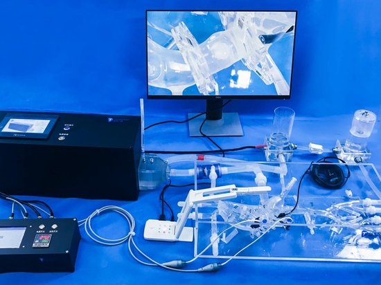
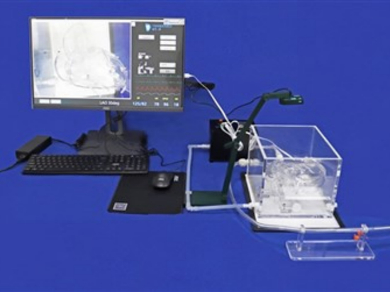
The Neural Intervention Training Suite II is a sophisticated simulation system crafted for neurovascular procedure training. At its core lies a neurovascular model that mirrors human blood circulation, paired with the Pulsatile Pump P-120 for lifelike pulsatile flow simulations. Complementing these are an advanced imaging setup furnished with DSA software for dynamic imaging, a visual presenter for lucid demonstrations, and an A4 lighting plate for heightened visualization. Bundled with vital accessories like simulated contrast agents, lubricants, thrombus materials, and necessary tools, this suite ensures a comprehensive training environment.
A novel feature of this suite is the incorporation of 3D printed models to elevate the training of medical practitioners in simulating intracranial angiography. These models offer a tactile and authentic portrayal of cerebral anatomy, empowering doctors to rehearse procedures meticulously and systematically. By integrating these cutting-edge models into training regimens, healthcare professionals can refine their abilities in navigating intricate neurovascular structures, comprehending pathological complexities, and mastering the nuances of performing angiographic processes with exactitude and assurance. This fusion of simulation technology not only enriches the educational journey but also fosters improved patient care outcomes by cultivating highly skilled healthcare providers.