#Industry News
What is transcatheter aortic valve replacement?
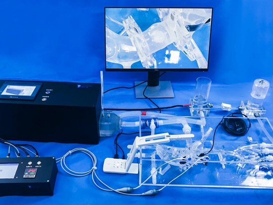
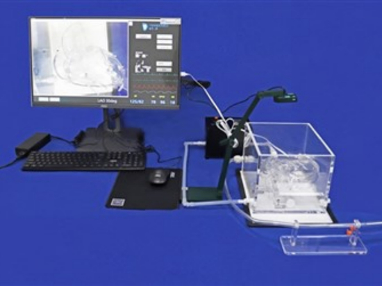
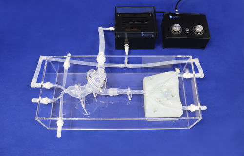
Model:Aortic Valve Model
Transcatheter aortic valve replacement (TAVR), also known as TAVR or transcatheter aortic valve implantation (TAVI), is a procedure to treat aortic stenosis, a narrowing of the aortic valve. The narrowing blocks the flow of blood to your body and forces your heart to work harder. This causes symptoms such as chest pain and shortness of breath.
TAVR replaces an aortic valve that is not working properly or is diseased with an aortic valve made from animal tissue. The TAVR procedure is minimally invasive, requiring only a small cut in the skin. It does not require open-heart surgery.The aortic valve is one of four valves that control blood flow in the heart. The aortic valve specifically controls the blood that runs from the heart through your aorta and to the rest of the body. Over time or because of a congenital heart defect, you can develop aortic stenosis, a type of heart valve disease.
There are several ways your doctor can perform TAVR, depending on your health and the health of your blood vessels.
●A TAVR using the femoral artery, a blood vessel in your groin or thigh, is the most common. Your doctor guides a tube with the replacement valve through the femoral artery to the heart.
●Blood vessels in the chest may be used by the doctor to guide the tube to your heart if your femoral artery is too small or damaged. This approach is called transapical access.
●Stomach area blood vessels may be used for TAVR if a patient’s leg arteries are too small or diseased for a more standard approach. NHLBI researchers developed this approach, called transcaval access, to make TAVR available to high-risk patients. It is less common. The doctor makes holes in both the vena cava, a major veins in your stomach area, and the nearby aorta to feed the tube with the replacement valve first through the vein and then through the aorta to the heart. You may be able to stay awake during this procedure. This type of TAVR approach may benefit women, who often have smaller blood vessels than men.
Accessing the heart from the vessel under the clavicle, or collar bone, may be an option if you have had heart surgery before or if you have another condition that makes it difficult to access other parts of the chest.
Your doctor may use the carotid artery in the neck to feed the tube to the heart — called transcarotid access. This type of procedure is rare but may be used when other options will not work.
Another rare location is through the septum, the wall of tissue that separates the right and left atria of the heart. Your doctor reaches the damaged valve by guiding the tube through a blood vessel from your thigh to the heart.