#Industry News
Some Knowledge of Atrial Septal Defect
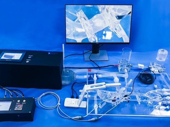
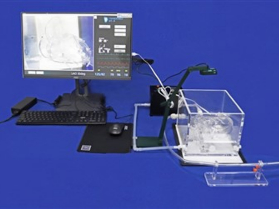
Model:Cardiac Vein With ASD
Atrial septal defect (ASD) is a congenital heart condition where there is an incomplete development of the septum between the left and right atria, leading to an abnormal passage between the two chambers. It is one of the common congenital heart diseases in children and accounts for 5%-10% of the incidence of congenital heart disease in our country.
Etiology and Pathogenesis
1.Etiology is related to intrauterine environmental factors during fetal development, maternal conditions, and genetic factors.
2.Classification: Atrial septal defects can be classified into primary defects and secondary defects. (1) Primary Defects: Located below the anterior part of the coronary sinus, with the lower edge of the defect close to the annulus of the mitral valve, often accompanied by a cleft in the mitral valve. (2) Secondary Defects: More common, located above the posterior part of the coronary sinus. The majority are single defects, with a few being multiple defects or fenestrations. Based on the anatomical location of the defect, they are further classified as central type (ostium secundum), superior type (sinus venosus type), inferior type, and mixed type. Secondary defects are often associated with other cardiac anomalies such as pulmonary valve stenosis and mitral valve stenosis.
Pathophysiology
In a normal heart, the pressure in the left atrium is higher than in the right atrium. With an atrial septal defect, blood from the left atrium shunts to the right atrium through the defect. The shunt volume depends on the pressure difference between the two atria, the size of the defect, and the resistance to filling of the left and right ventricles. In newborn infants, the pressures in the two atria are similar, and there is minimal shunting through the defect. As age increases, the pressure difference between the atria increases, leading to increased shunting from left to right, often reaching 2 to 4 times the systemic blood flow.
The increased volume load on the right heart leads to enlargement of the right atrium, right ventricle, and pulmonary artery. The increased blood flow in the pulmonary circulation raises pulmonary artery pressure, leading to reactive constriction of the pulmonary arterioles. Long-term constriction causes intimal thickening, medial hypertrophy, fibrosis, luminal narrowing, increased pulmonary vascular resistance, ultimately resulting in obstructive pulmonary arterial hypertension. When the pressure in the right atrium exceeds that in the left atrium, right-to-left shunting occurs, causing cyanosis, Eisenmenger syndrome, and eventually death due to right heart failure.
Primary defects with accompanying mitral valve clefts lead to mitral regurgitation, further increasing the left-to-right shunt volume. Pulmonary arterial hypertension occurs earlier in these cases, with more pronounced pathological and clinical progression compared to secondary defects.
Clinical Symptoms:
1.Symptoms: Patients with small shunts in secondary atrial septal defects may be asymptomatic in childhood and are often detected during routine physical examinations. Symptoms such as exertional dyspnea, fatigue, palpitations, respiratory infections, and right heart failure typically appear in young adulthood. Patients with primary atrial septal defects and severe mitral regurgitation may present early with symptoms of heart failure and pulmonary arterial hypertension. Severe pulmonary arterial hypertension can lead to right-to-left shunting and cyanosis.
2. Signs: (1) Inspection: Primary atrial septal defects result in significant cardiac enlargement and prominence in the precordial area. Secondary defects may present with cyanosis and clubbing of fingers/toes. (2) Palpation: There may be a lift in the precordial area, with rare cases exhibiting palpable thrills. (3) Auscultation: In the pulmonary valve area, a grade II-III blowing systolic murmur may be heard, along with accentuated second heart sound and fixed splitting. In cases with large shunts, a soft diastolic murmur may be audible at the cardiac apex. In patients with pulmonary arterial hypertension, the systolic murmur in the pulmonary valve area diminishes, the second heart sound becomes more prominent and split.
The Cardiac Vein With ASD I model is a sophisticated medical training tool designed to simulate various cardiovascular procedures and conditions. This model features an array of veins including the internal jugular vein, external jugular vein, SVC, IVC, subclavian vein, cephalic vein, basilic vein, brachial vein, median cubital vein, as well as critical cardiac structures such as the left and right atrium, pulmonary vein, renal vein, iliac vein, and femoral vein. Notably, this model incorporates an atrial septal defect (ASD) with an internal diameter of 8mm within the atrial septum, providing a realistic representation of this congenital heart anomaly. The design of the model includes access ports in the femoral vein, median cubital vein, cephalic vein, and basilic vein, allowing for the insertion of devices for procedures like PICC line insertion, atrial septal puncture, and pulmonary vein ablation.
This model serves as a versatile tool for training, development, testing, and demonstration of various cardiovascular interventions. It is particularly useful for simulating procedures such as atrial septal puncture and pulmonary vein ablation. Medical professionals can utilize this model to practice and enhance their skills in these procedures in a controlled and realistic setting. Furthermore, the customization options offered for this model allow for tailoring its features to specific requirements, including adjusting the size of the atrial septal defect, the complexity of the pulmonary vein and IVC structures, and even creating custom models based on provided data files in various formats. With its detailed representation of cardiac and vascular anatomy, along with the simulated ASD, this model offers a valuable resource for medical training, device testing, and educational purposes in the field of cardiovascular medicine.