#Product Trends
New perspectives on ultrasound imaging
3D reconstruction, image fusion, and precise localization
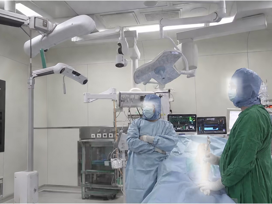
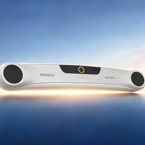
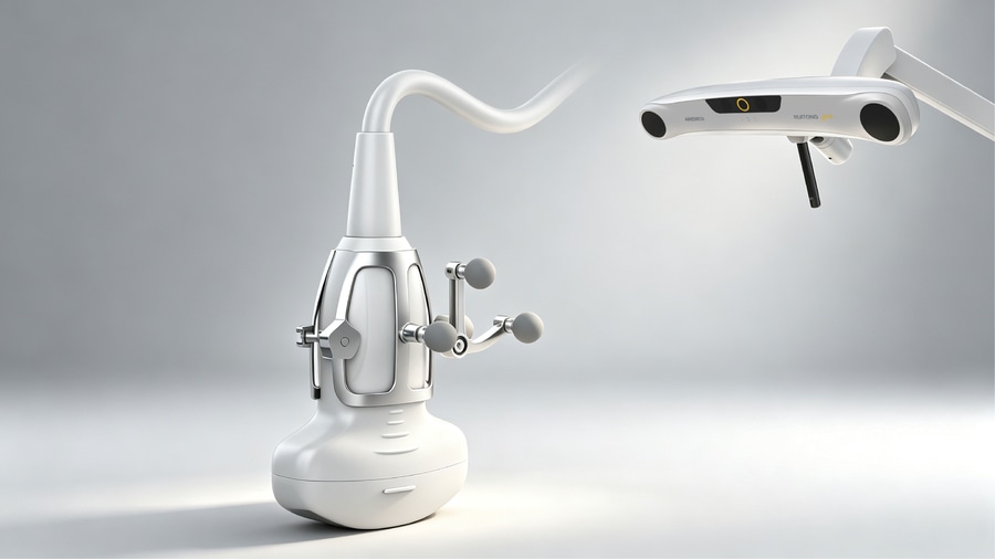
Ultrasound, with its advantages of real-time, convenient, and radiation-free, is widely used in organ tissue puncture biopsy, ablation therapy, catheterization and drainage, and drug injection, providing safety assurance for surgical procedures. To address the limitations of traditional two-dimensional ultrasound planar imaging, the platform relies on the AirMeddy Ruitong optical positioning camera and Ruixing magnetic positioning sensor to achieve precise spatial posture tracking, integrating ultrasound 3D reconstruction, multimodal image fusion, and spatial positioning technologies to broaden the application boundaries of ultrasound diagnosis and treatment and interventional scenarios.
1. Three-dimensional ultrasonic reconstruction: from two-dimensional cross-sections to stereoscopic views
Typically, two-dimensional ultrasound can only output a single facet, making it difficult to fully present the overall morphology and stereoanatomy of organs and tissues. 3D ultrasonic reconstruction relies on positioning sensors to achieve precise spatial stitching of multi-faceted images. In clinical applications, markers can be fixed and tracked on ultrasound probes to pre-calibrate the transformation relationship between the ultrasound imaging plane and the marker coordinate system.
During surgery, the AirMeddy Ruitong optical positioning system or Ruixing magnetic positioning equipment can be used to track or sense markers on the ultrasound probe in real time to obtain probe displacement and angle information, and to calculate the position and posture of each ultrasound image frame in real time. After spatial mapping, the dispersed two-dimensional planes are fused into a unified three-dimensional reference frame, achieving precise image sequence stitching and 3D reconstruction. Paired with intelligent segmentation algorithms, it can accurately distinguish lesions, blood vessels, and normal tissue, visually present anatomical adjacency relationships, support quantitative calculation of lesion volume and size, and help improve missed diagnoses and misjudgments common in two-dimensional sections.
AirMedy - Embodied Intelligent Navigation Platform Single Page Mobile Version - 14(1) .jpg
Four-dimensional ultrasound superimposed with time dimensions, relying on optical/magnetic positioning sensors, can capture organ displacement caused by breathing in real time, effectively avoiding the false positioning caused by the two-dimensional partial volume effect, reducing deviations, intuitively presenting the three-dimensional spatial relationship between surgical instruments and lesions, supporting real-time adjustment of instrument direction and depth during surgery, and adapting to dynamic guidance needs.
Screenshot 161901.png 2026-07-27
2. Ultrasound-CT fusion: a technological path with complementary advantages
The core purpose of integrating ultrasound and CT is to achieve complementary advantages. Real-time ultrasound captures dynamic information during surgery, such as organ movement and changes in the position of surgical instruments; With high-precision CT, a detailed anatomical base map is provided, clearly presenting the structure of the tissue and the location of lesions. The spatial alignment and real-time fusion of the two is like giving doctors a pair of "fluoroscopy eyes," allowing them to clearly see anatomical details and track instrument positions in real time, significantly improving the accuracy and safety of procedures such as puncture, ablation, and biopsy.
Screenshot 161908.png 2026-07-27
3. Spatial positioning technology: the core support of navigation systems
Precise spatial positioning is the foundation of ultrasound-guided navigation systems. By utilizing external tracking systems, 3D coordinates of probes and instruments are obtained, providing unified spatial references for 3D reconstruction, image registration, and intraoperative navigation. Optical and electromagnetic positioning technologies are mainstream in clinical practice.
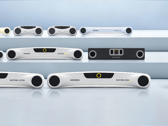
Optomagnetic series .jpg
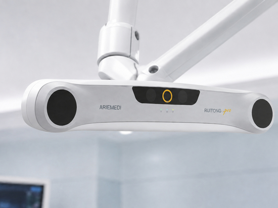
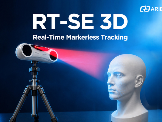
iMedic independently developed high-precision medical-grade Ruitong optical positioning cameras and Ruixing magnetic positioning devices, breaking the long-standing overseas monopoly and providing a precise spatial positioning foundation for 3D ultrasonic fusion. Optical navigation accuracy reaches sub-millimeter level, ensuring coordinate consistency across multiple facets. Magnetic navigation without obstruction, magnetic field penetrates soft tissue, suitable for deep intervention scenarios. The two can work independently or collaboratively, providing stable real-time six-degree-of-freedom posture tracking for ultrasound probes and instruments, improving navigation efficiency, and suitable for minimally invasive surgery, interventional treatments, and other clinical scenarios requiring high-precision spatial positioning.
New ultrasound imaging relies on optical or magnetic positioning sensor guidance, integrating 3D reconstruction, image fusion, and spatial positioning technologies, effectively overcoming the inherent limitations of 2D ultrasound. It can be expanded to routine examinations, clinical diagnosis and treatment, and minimally invasive interventions, improving lesion detection rates, achieving precise localization, and reducing the dependence on the physician's personal experience.
——Contact Us——
Phone: 400-660-3568 (ext. 2) / 13001067056
Email: [email protected]