#Product Trends
Precision Navigation for Non-Invasive Deep-Brain Targeting
Ruitong Stereo Camera Opens New Possibilities for Focused Ultrasound
Traditional non-invasive brain stimulation techniques have long been limited by two major challenges: low spatial resolution and insufficient penetration depth. Transcranial focused ultrasound (tFUS) offers a promising new solution.
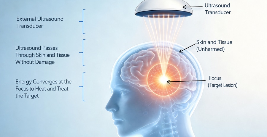
By delivering low-intensity focused ultrasound through the skull, tFUS enables non-invasive targeting of deep-brain structures, overcoming the limited penetration and poor focal precision of conventional stimulation methods. It can both excite and inhibit neural activity and reach deep regions such as the hippocampus and thalamus. Its neuromodulatory effects are believed to involve the activation of mechanosensitive ion channels in neuronal membranes, enabling millimeter-level, point-specific targeting.
A navigated transcranial ultrasound stimulation system consists of two primary modules: an ultrasound stimulation module and a navigation module. Within the navigation module, the optical stereo-tracking camera acts as the system’s “spatial awareness center.” Through high-frame-rate, high-precision optical tracking, it provides the accurate spatial positioning required for millimeter-level targeting.
01 See the Target Clearly: Navigation for Non-Invasive Focused Ultrasound
Improving the accuracy of a navigated transcranial focused ultrasound system depends on the system’s ability to capture the spatial position and orientation of both the ultrasound transducer and the patient’s head in real time—and to dynamically compensate for movement.
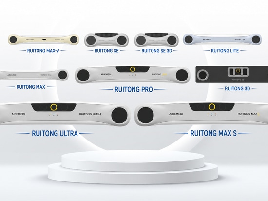
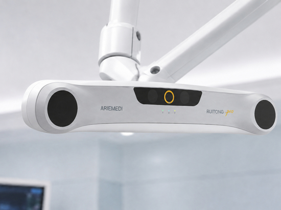
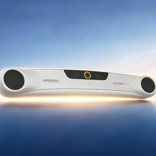
ARIEMEDI’s independently developed Ruitong PRO optical tracking camera delivers an RMS positioning accuracy of 0.12 mm, together with a wide field of view and high frame rate. It combines precision positioning, real-time tracking, 3D reconstruction, and dynamic motion compensation in a single system.
The camera continuously tracks optical markers attached to the ultrasound transducer and calculates its six-degree-of-freedom position and orientation in real time. With one-click scanning, it can acquire millions of point-cloud data points to register the MRI-derived anatomical model with the patient’s physical position.
It can also detect head movement and respiratory motion almost instantly, providing real-time positional feedback to a mechanical compensation system and helping reduce the risk of the ultrasound focus drifting away from the intended target.
02 Precise, Real-Time, and Efficient: Key Advantages of Ruitong Optical Tracking for Focused Ultrasound Navigation
Precise and Non-Invasive Targeting
A focused ultrasound navigation system can integrate MRI data to create a patient-specific 3D model of the brain. The Ruitong optical tracking system monitors the ultrasound transducer’s 3D coordinates with submillimeter precision and accurately determines its position and orientation relative to the patient’s head.
This helps ensure that the ultrasound focus reaches the intended brain region while reducing exposure to non-target areas. Such precision is particularly important when targeting deep-brain nuclei or anatomically variable lesions associated with conditions such as epilepsy and disorders of consciousness.
Real-Time Targeting
The Ruitong optical navigation system can visualize the ultrasound beam path and target location, helping operators align the focal point with the intended brain region and minimize the effects of side lobes and unintended exposure.
Even when the ultrasound transducer is moved over a relatively large area, the system continuously tracks its optical markers and compensates for positional deviations in real time, helping maintain stable and continuous stimulation.
Greater Workflow Efficiency
Traditional manual landmark registration and standardized head-model matching can be time-consuming and may not adequately account for anatomical differences between patients.
By integrating optical tracking with 3D reconstruction, the Ruitong stereo camera can rapidly register the ultrasound transducer, the patient’s head, and the medical imaging model within seconds. Compared with conventional workflows, this can substantially reduce preparation time, minimize repeated trial positioning, and improve patient comfort—particularly for children, older adults, and patients with movement disorders.
03 From Precision Positioning to Precision Intervention: Applications and Future Potential
With the stable spatial reference provided by the Ruitong camera, navigated transcranial ultrasound systems are showing promise across a growing range of clinical research areas:
Alzheimer’s disease: Targeting the hippocampus and default mode network to investigate potential improvements in cognitive function.
Epilepsy: Targeting the thalamus or regions surrounding an epileptic focus to explore the potential reduction of seizure frequency.
Disorders of consciousness: Stimulating the thalamus or brainstem reticular activating system to support research into recovery of consciousness.
Psychiatric conditions: Modulating the prefrontal cortex or limbic system, with early studies reporting encouraging signals in depression and anxiety.
The positioning accuracy and smooth workflow enabled by stereo vision can improve more than a single treatment session. It also provides a reproducible spatial reference for repeated treatment, multimodal data acquisition, and the development of individualized treatment protocols.
Navigated transcranial ultrasound stimulation combines the non-invasive advantages of low-intensity focused ultrasound with the millimeter-level accuracy of image-guided navigation.
At the heart of this integration is the Ruitong optical stereo-tracking camera—the system’s “precision eye.” By providing accurate, reliable, and real-time 3D spatial data, it supports the transition of deep-brain focused ultrasound navigation from approximate positioning to precise, target-specific intervention.