#Product Trends
Cardiac Ultrasound, Demystified: How AI Labeling Is Bringing the Heart Into Primary Care
For clinicians outside cardiology, interpreting a cardiac ultrasound image has always been the hard part. A new AI-powered overlay changes that — labeling every structure, in real time, as you scan.
Point-of-care ultrasound has steadily expanded beyond the emergency department, but cardiac imaging has remained one of its more intimidating frontiers. The issue is rarely the scan itself — it is knowing what you are looking at. Four chambers, two valves, a dynamic image that shifts with every millimeter of probe movement: for a primary care clinician just building their POCUS skills, the gap between acquiring an image and confidently reading it can feel wide.
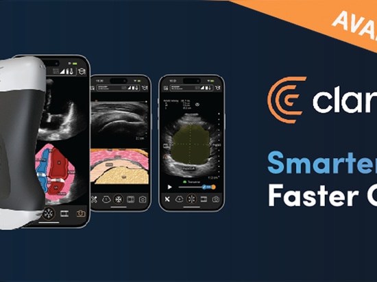
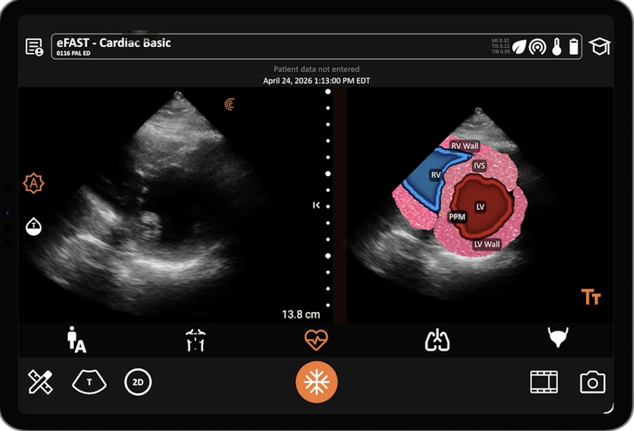
Clarius T-Mode™ for the Heart was designed specifically to close that gap. Built into the Clarius PAL HD3 wireless ultrasound scanner, T-Mode overlays real-time anatomical labels directly onto the cardiac image as the clinician scans — identifying the left ventricle, right ventricle, left atrium, aortic outflow tract, and mitral valve without any manual input. As the probe angle changes, the labels adjust instantly, keeping pace with the image.
In a classroom demonstration led by Dr. Tatiana Havryliuk — emergency physician and founder of Hello Sono, a program supporting compliant focused ultrasound programs in medical practices — the feature is shown across three standard cardiac views: parasternal long axis, parasternal short axis, and apical four chamber. In each, T-Mode identifies the relevant structures automatically, turning what can feel like an ambiguous grey-scale image into a clearly annotated, readable snapshot of the heart.
The clinical implications for primary care are significant. Cardiac POCUS offers fast, meaningful clinical data — ventricular function at a glance, pericardial effusion, gross valve assessment — that can guide triage, referral decisions, and immediate management. The barrier has not been access to the technology; it has been confidence in interpretation. By making anatomy legible from the first scan, T-Mode lowers that barrier meaningfully, accelerating the learning curve for novice users while reinforcing anatomical recall for those building volume.
As wireless, handheld ultrasound continues to expand across primary care settings, tools that reduce the cognitive load of interpretation — rather than adding to it — will define which platforms clinicians actually reach for. T-Mode is a clear step in that direction.