#Industry News
Tow-Int WBP in the study of opioid morphine withdrawal
The TOW-INT WBP has assisted Academician Ma Lan's team in making significant advancements in the study of opioid morphine withdrawal
Introduction to the Research
The abuse of opioid drugs poses a significant threat to economic development, social stability, and public health. Morphine, a commonly used opioid analgesic in clinical practice, is limited in its application due to side effects such as addiction and analgesic tolerance. Opioid addiction is a complex brain disorder, where prolonged and repeated exposure to addictive substances causes neuroadaptive changes in the brain, leading to euphoria (positive reinforcement). Additionally, after discontinuation of the addictive drug, users experience severe withdrawal symptoms and negative emotions (negative reinforcement). To avoid the negative emotions and withdrawal symptoms caused by drug cessation, drug-seeking behavior is further reinforced. The alternating cycles of positive and negative reinforcement are critical factors in drug abuse and relapse among addicts. Therefore, understanding the mechanisms underlying negative reinforcement triggered by morphine withdrawal could help alleviate withdrawal-induced negative emotions, providing new intervention targets to prevent addiction relapse and drug abuse, making this research of significant scientific and societal importance.
Research Methodology
On January 18, 2024, Academician Ma Lan's team from the Institute of Brain Science/Frontiers Science Center for Brain Science at Fudan University published a research paper titled "Targeting mitochondrial dynamics of morphine-responsive dopaminergic neurons ameliorates opiate withdrawal" in the Journal of Clinical Investigation. For the first time, the study analyzed the role of morphine-activated dopaminergic neuron clusters in the ventral tegmental area (VTA) in negative reinforcement and their molecular and cellular mechanisms, from the perspective of mitochondrial homeostasis. The study discovered that inhibiting mitochondrial fission with Mdivi-1 or overexpressing the mitochondrial fusion gene Mfn1 blocked mitochondrial fragmentation in dopaminergic neurons following long-term morphine administration and alleviated pathological changes in neuronal plasticity, thus reducing the negative reinforcement and analgesic tolerance side effects of morphine. During morphine-based analgesic treatments, tolerance, respiratory depression, constipation, and other side effects often occur. The team found that Mdivi-1 effectively mitigates morphine-induced analgesic tolerance following long-term use. Using mouse volumetric imaging to monitor respiratory parameters, the study demonstrated that Mdivi-1 treatment could reduce damage to the mitochondrial respiratory chain in the VTA caused by morphine, prevent the downregulation of activity in dopaminergic neuron clusters, and alleviate mitochondrial fragmentation, ultimately mitigating withdrawal symptoms, negative emotions triggered by morphine withdrawal, and long-term drug craving, providing new intervention strategies and targets for morphine dependence.
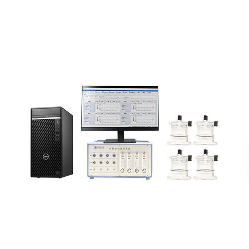
Mouse Whole-body Plethysmography
The whole-body plethysmography (TOW-INT TECH) was used to measure respiratory data. It monitored the respiratory rate, tidal volume, and peak flow of unrestrained mice. Airflow sensors were connected to each chamber’s port to maintain a stable airflow. Before the experiment, each chamber was calibrated with its connected sensor. The mice were acclimated to the test chambers for 30 minutes, and respiratory parameters were recorded for 10 minutes to establish baseline data before administering saline or morphine (10 mg/kg). 45 minutes before morphine injection, the mice were pre-treated with a vehicle or Mdivi-1 (50 mg/kg). Respiratory parameters were then collected 90 minutes after morphine administration. Respiratory inhibition was assessed using the mouse volumetric imaging method. Morphine administration (10 mg/kg, intraperitoneal) caused a significant decrease in respiratory rate 15 minutes after injection. Statistical analysis using RM ANOVA with Bonferroni tests showed significant differences between saline and morphine groups (n=4 mice/group) and between morphine+vehicle and morphine+Mdivi-1 groups (n=8 mice/group).
Research Results
The reduced activity of dopaminergic, morphine-responsive neurons in the VTA mediates conditioned aversion and anxiety during morphine withdrawal. The plasticity of VTA neurons is critical for the occurrence of morphine withdrawal. The neuron cluster that responds to initial morphine exposure (Mor-Ens) in the VTA preferentially projects to the nucleus accumbens (NAc) and induces dopamine-dependent positive reinforcement. To investigate maladaptive changes in dopaminergic Mor-Ens, Cre-Loxp and Flpo-Frt-dependent robust activity marking (RAM) systems were used to label jGCaMP7b in these dopaminergic Mor-Ens without doxycycline (Dox). AAV-RAM-TTA-TRE-flpo, AAV-fDIO-TH-Cre, and AAV-DIO-jGCaMP7b viruses were delivered, and optical fibers were unilaterally implanted in the VTA. Spontaneous Ca2+ events were recorded before and after escalating morphine administration (morphine EDA) in the home cage. Ca2+ event frequency decreased after morphine EDA (day 7) compared to baseline (day 1), while saline treatment had no effect. Immunostaining revealed that most GCaMP7b+ cells were confined to the VTA and co-labeled with tyrosine hydroxylase (TH). These data indicate that spontaneous neuronal activity in VTA dopaminergic Mor-Ens decreases following morphine EDA.
Research Conclusion
This study reveals that mitochondrial dysfunction in the brain's dopamine system is a critical cellular mechanism for the negative reinforcement induced by long-term opioid use. It suggests that mitochondrial dynamics could be a novel therapeutic target for opioid use disorder. Mitochondrial-targeted drugs may hold significant translational value in the treatment of opioid addiction.
References
https://www.jci.org/articles/view/171995
https://iobs.fudan.edu.cn/1a/16/c17247a662038/page.htm
http://k.sina.com.cn/article_5803416260_159e91ac4001013pbh.html
Additional references: http://www.tow-int.com/index.php/CLIENT1/
TOW-INT TECH Provides WBP Solutions
Whole-body Plethysmography System
The whole-body plethysmography (WBP) system developed by TOW-INT TECH allows the measurement of respiratory parameters in conscious and freely moving animals, such as respiratory rate, tidal volume, and airway hyperresponsiveness (AHR) testing. During testing, the animals remain conscious and unrestrained, avoiding the need for invasive tracheotomy or anesthesia, making the experimental process more straightforward. This system is used for studying the responsiveness of respiratory system model animals to drugs, as well as for pharmacological and toxicological studies of respiratory medications. It is particularly suitable for rapid multichannel screening in large groups of animals, long-term tracking studies, and repetitive screening tests.
Contact us now!
We are committed to making your research easier, more accurate, and more efficient and helping you build confidence in your data! We have provided services for a large number of customers, giving us rich experiences in offering customized, professional solutions according to your needs.