#Industry News
Regulation of respiratory rhythms by brain neural circuits to combat anxiety
WBP combined with optogenetics
Have you ever wondered if breathing is merely an instinctive response of the body, or if it is intricately connected to our emotions and behaviors?
A research team led by the Salk Institute for Biological Studies in the United States published a groundbreaking study in Nature Neuroscience, which partially reveals how the cerebral cortex precisely controls breathing, particularly how the top-down respiratory circuit influences the brainstem network.
WBP combined with optogenetics
Research Background
Breathing, a seemingly simple physiological activity, is much more than just maintaining gas balance. Its rhythm can be influenced by various behaviors, such as speaking, swallowing, and even emotional changes, which can alter the breathing pattern. Humans have the ability to consciously control their breathing rhythm, and many cultures practice slow breathing or mindfulness techniques to regulate emotions. However, how the brain’s cortex precisely controls breathing—especially the top-down control of respiratory circuits on the brainstem network—remains a mystery.
Research findings have identified a neural circuit that slows down breathing through extensive studies in mice. This circuit, which projects from the dorsal anterior cingulate cortex (dACC) to the pontine reticular nucleus caudal (PnC), was found to reduce the respiratory rate. Using optogenetic techniques to activate this cortical-pontine pathway (dACC → PnC neurons), the mice’s breathing significantly slowed, and anxiety-related behaviors were alleviated, although emotional valence did not change. Further research revealed that dACC neurons project to inhibitory GABAergic neurons in the PnC, which, in turn, project to the brainstem’s respiratory center, regulating the breathing rhythm. This suggests that the input from the prefrontal cortex (dACC/M2) to the PnC forms a top-down regulatory circuit inducing slow breathing.
The correlation between neuronal activity and behavioral breathing changes was observed in various behaviors such as drinking, swimming, vocalizing, and sniffing. For example, during drinking, dACC → PnC neurons were briefly activated, and the activity of downstream PnC GABAergic neurons significantly increased, coordinating breathing with swallowing. In swimming tests, different swimming conditions led to variations in breathing patterns and neuronal activity, but all showed a correlation between neuronal activity and respiratory cycles. In vocalizing behaviors, such as foot-shock-induced vocalization, the activity of dACC → PnC neurons was also linked to changes in breathing. These results suggest that, although many behaviors are controlled by somatic and trigeminal reflexes, the top-down slow breathing pathway also plays a crucial role in respiratory regulation.
dACC→PnC Activity and Emotion-Induced Breathing Changes
In anxiety-provoking environments, such as the elevated plus maze test, the breathing and behavior changes of mice were closely associated with dACC → PnC neuronal activity. In the exposed area, the mice exhibited faster breathing, whereas when they “escaped” to the enclosed area, their breathing slowed, and dACC → PnC neuronal activity increased. In open-arm exploration, neuronal activity correlated with the mice’s behavior: activity persisted or increased when the exploration was successful, and decreased when unsuccessful. In another unavoidable anxiety-inducing experiment, similar changes in breathing and neuronal activity were observed. These results suggest that dACC → PnC neuronal activity is associated with breathing and behavior in anxiety-inducing environments, and increased neuronal activity may facilitate anxiety relief.
dACC→PnC Neural Circuit in Alleviating Anxiety-like Behaviors
Optogenetic activation of the dACC → PnC neurons was found to inhibit anxiety-like behaviors in mice and promote exploratory behavior, without altering emotional valence or approach behaviors. For example, in real-time place preference and female odor preference experiments, optogenetic stimulation did not alter mouse behavior, while in aversive environment tests, such as the fox feces odor (TMT) test, optogenetic stimulation eliminated the mice’s avoidance behavior. Further studies indicated that the projection of dACC → PnC neurons is crucial for controlling anxiety-related responses. The downstream PnC GABAergic neurons, projecting to the respiratory centers and forebrain areas related to fear and anxiety, may regulate both breathing and emotion, thus alleviating anxiety.
Importance of dACC→PnC Neurons in Coordinating Breathing and Behavior
Inhibition of the dACC → PnC circuit affects the coordination of behavior and breathing in mice. In the drinking experiment, inhibition of this circuit decreased the success rate of drinking and shortened the breathing cycle, indicating that this circuit is crucial for coordinating slow breathing and drinking behavior. In anxiety-related behavioral tests, such as the light/dark choice model, inhibition of this circuit reduced the time spent in the light area and decreased the proportion of slow breathing cycles, further confirming the necessity of the dACC → PnC circuit in coordinating behavior and inducing slow breathing to alleviate anxiety.
Research Significance
This study significantly advances our understanding of top-down control of breathing and reveals the neural circuit mechanism through which slow breathing and anxiety relief are jointly regulated. This not only helps us better understand how the brain regulates breathing and emotion but also provides potential new targets for the treatment of anxiety disorders and related conditions. In the future, we may be able to modulate this neural circuit to develop more effective therapeutic approaches to help people better manage emotions and cope with stress.
Experimental Methods
Animal Experiments
All experiments were approved by the Institutional Animal Care and Use Committee (IACUC). Wild-type or Vgat-ires-Cre transgenic mice were housed under specific conditions for various experiments.
Breathing Measurement
Breathing was monitored using inductance plethysmography (under anesthesia) and nasal thermistor (during awake behavioral states).
Stereotaxic Surgery
Viruses or tracers were injected into specific brain regions to perform optogenetic control, calcium activity monitoring, and other operations.
Behavioral Experiments
These included drinking, swimming, vocalizing, sniffing, elevated plus maze tests, and other experiments. Behavioral and neuronal activity were monitored using optogenetic stimulation or fiber photometry.
Experimental Results
Inhibition of the dACC → PnC circuit affected drinking behavior, resulting in a lower success rate of drinking and a shorter breathing cycle, indicating that this neural circuit is essential for coordinating drinking and breathing. In the light/dark choice test, inhibition of the dACC → PnC circuit reduced the time spent in the light zone and decreased the proportion of slow breathing cycles, indicating its necessity for inducing slow breathing to alleviate anxiety.
Optogenetic activation of the dACC → PnC neurons did not alter the behavior of mice in real-time place preference and female odor preference tests, but it alleviated their avoidance response to aversive stimuli, such as the fox feces odor (TMT), and increased exploratory behavior in tests like the elevated plus maze.
Stimulation of the dACC → PnC neurons, which project to the PnC axon terminals, increased exploratory behavior, while inhibition decreased it, indicating the importance of these projections in controlling anxiety-related responses.
The PnCGABA neuron axons project to brain areas associated with both respiration and anxiety, suggesting that slow breathing may alleviate anxiety through these connections.
In the elevated plus maze experiment, mice exhibited faster breathing in the exposed area, and their breathing slowed when they “escaped.” The activity of dACC → PnC neurons increased during escape, and their activity was associated with both breathing and anxiety-related behaviors.
In a similar but unavoidable anxiety-provoking environment, the mice’s breathing accelerated, and the activity of dACC → PnC neurons decreased.
During drinking, the dACC → PnC neurons and PnCGABA neurons were briefly activated when the mice ingested water, coordinating with the respiratory cycle. During swimming, variations in breathing patterns and neuronal activity were observed under different testing conditions, indicating that the top-down pathway participates in regulating breathing rhythms during swimming.
In vocalizing, dACC → PnC neuronal activity was associated with slow expiration cycles; during sniffing, dACC → PnC neuronal activity was linked to changes in breathing cycles, showing that their activity is influenced by behavior.
Brain mapping identified the PnC as a possible region, and retrograde tracing revealed that the dACC → PnC projection neurons (dACC → PnC neurons) are mainly located in layer 5 of the dACC and the secondary motor cortex (M2).
Optogenetic activation of dACC → PnC neurons reduced the breathing frequency in anesthetized mice, but this effect was absent in deeper anesthesia, suggesting that the circuit operates under certain conditions.
Anterograde tracing and immunostaining confirmed that dACC neurons project to GABAergic inhibitory neurons in the PnC (PnCGABA neurons), and optogenetic activation or inhibition of PnCGABA neurons could slow down or accelerate breathing frequency.
Experimental Conclusion
A top-down brain circuit, the dACC → PnC circuit and its downstream PnCGABA neurons, was identified, which promotes slow breathing cycles and responds to behavioral modulation of breathing.
Activation of this circuit can alleviate anxiety-like behaviors and is crucial for coordinating breathing and behavior. These findings provide new insights into the neural mechanisms of breathing control and emotion regulation.
Research Discussion
Optogenetic Techniques for Treating Mood Disorders
Optogenetic techniques can be applied to treat mood disorders. Integrating knowledge from optics, software control, genetic manipulation, and electrophysiology, optogenetics offers unique high spatiotemporal resolution and cell-type specificity, enabling precise, non-invasive stimulation of neurons. By stimulating specific neuronal groups, optogenetics holds promise for modulating neural circuits related to emotions, potentially improving symptoms of mood disorders. Although clinical applications of optogenetic techniques for treating mood disorders are still under exploration, this technology offers new directions and ideas for mood disorder treatments.
Tow-Int Optogenetic version WBP
Optogenetic function WBP
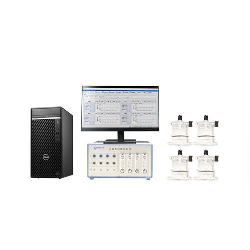
Tow-Int Tech Whole-Body Plethysmography System
The system utilizes unrestrained whole-body plethysmography to perform pulmonary function and airway responsiveness tests on awake, freely moving small animals. The respiratory movements of the animals cause changes in the thoracic volume within the plethysmograph chamber. These volume changes are converted into electrical signals through a pressure transducer and amplifier, which are then processed by a computer to display the respiratory curve and calculate various respiratory parameters, such as tidal volume (TV), peak expiratory flow (PEF), and respiratory rate.
Optogenetic Collaborative Research: Optogenetics is combined to explore the mechanisms of respiratory rhythm control.
Contact us now!
We are committed to making your research easier, more accurate, and more efficient and helping you build confidence in your data! We have provided services for a large number of customers and have rich experiences in offering customized, professional solutions according to your needs.