#Industry News
What is Bronchoscopy, What to expect when undergoing this test?
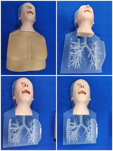
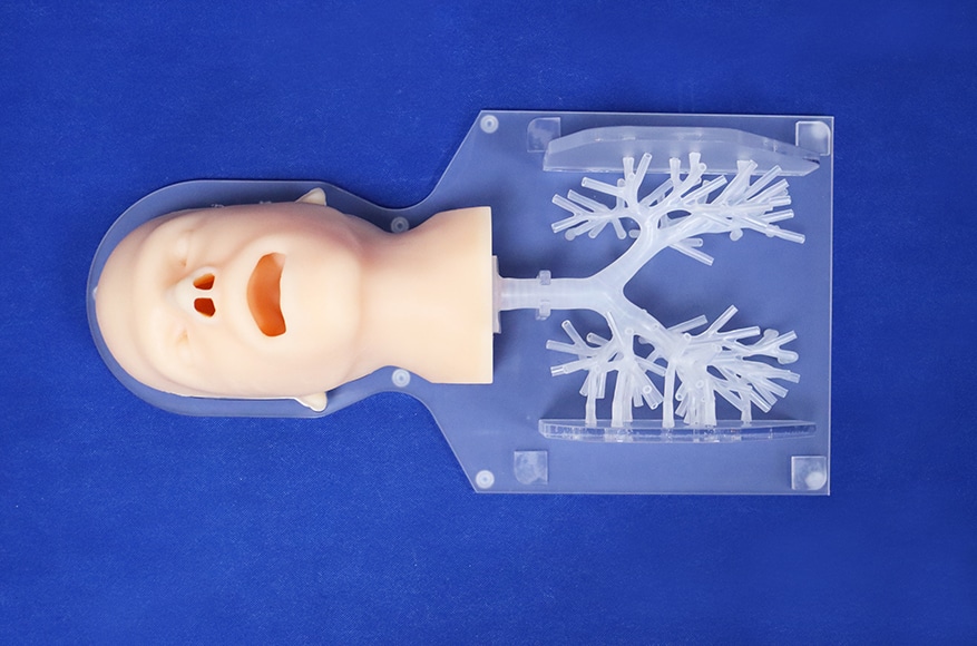
Bronchoscopy Simulator II
A bronchoscopy is an endoscopic medical procedure that is used to look inside the airways (bronchi) and the lungs. It involves inserting a bronchoscope—a narrow tube that has a light and a camera on one end—through the nose or mouth and guiding it down through the trachea (windpipe) in order to get an internal view of the respiratory system. It may be done to diagnose a disease or condition such as lung cancer or infection, or to treat a medical problem such as a foreign object that's lodged in the airways.
There are two main reasons a person might need a bronchoscopy:
Evaluation
Your healthcare provider may opt to perform a bronchoscopy to evaluate symptoms and other indications that something may be wrong with the lungs or airways. Examples include:
A chronic cough—one that has lasted for more than three months with no obvious cause
Hemoptysis (coughing up blood)
Shortness of breath or low oxygen levels
A suspicion there may be something lodged in your airways
An imaging test that showed a tumor or growth on a lung, scarring or other changes to the lung tissue, or the collapse of an area of a lung
Symptoms of infection in the lungs or bronchi that can't be diagnosed another way or require a special type of evaluation
Signs of rejection after a lung transplant
Inhalation of a toxic gas or chemical
A bronchoscopy also can be used to take a biopsy of abnormal lung or airway tissue, to biopsy the lymph nodes in the central chest adjacent to airways for evidence of cancer involvement, and to visualize tumors within the lungs that do not extend into the bronchi using a technique known as endobronchial ultrasound (EBUS).
In this procedure, a tumor deep in the airways may be visualized with ultrasound and biopsied during a bronchoscopy (an ultrasound-guided needle biopsy). EBUS can also be used to obtain a sample from lymph nodes that are adjacent to the airways.
In addition to techniques designed to look deeper than the airways during a bronchoscopy, there are also several new technologies used to diagnose early lung cancers. These include radial EBUS bronchosopy, robotic assisted bronchoscopy, narrowband imagery, and high magnification video bronchoscopy.
Treatment
By providing both access and a direct view of the inside of the airways and lungs, a bronchoscopy can allow a healthcare provider to perform all sorts of treatments, such as:
Removing fluid or mucus from airways
Removing a foreign object from airways
Widening (dilating) an airway that is blocked or narrowed
Washing out an airway
Bronchoscopy can also be used as part of certain treatments for lung cancer that is in or near the large airways. It might be used to assist with a procedure called brachytherapy, for instance, in which radiation is delivered directly to a tumor through the bronchoscope.
There are two types of bronchoscopy. The most common uses only a flexible bronchoscope and requires on local anesthesia and a light sedative; sometimes general anesthesia is necessary, depending on the length of the procedure. Less often a rigid bronchoscope, which is thicker than a flexible one and typically made of metal, is necessary. In that case, a patient must be under general anesthesia in an operating room.
Which one your healthcare provider opts for will depend on the purpose of the test and your overall condition.
Risks and Contraindications
Most people tolerate both types of bronchoscopy quite well. There are some potential risks, however.Though they are not common, they include:
Spasms in the airway such as laryngospasm (spasm of the larynx) or bronchospasm (spasm of the bronchi)Cardiac complications such as an abnormal heart rhythm or heart attack in people with existing heart disease
Low blood oxygen
Breathing difficulties
Pneumothorax (a collapsed lung): This can happen if the lung is punctured during the procedure, allowing air to collect in the space around the lungs. If small, your healthcare provider may simply follow it with a chest X-ray. If it is large, a chest tube may need to be placed to remove the air and you may need to be admitted to the hospital.