#Industry News
What is the diagnosis and treatment of aortic diseases?
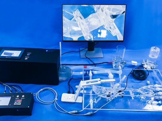
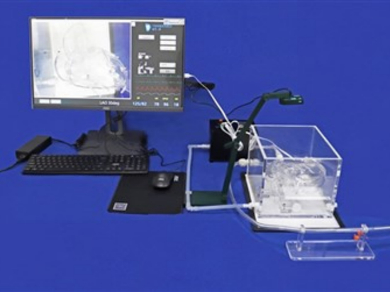
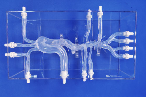
Model:Aortic Dissection(XXK004D)
What is the aorta?
The aorta is referred to as the "life's golden cane." It originates from the heart and extends to the iliac arteries, resembling a "cane." The branches of the aorta supply blood to various organs in the body, ensuring the normal functioning of the human body. Any pathology occurring in any part of this "cane" can affect the blood supply to various organs, posing a threat to life. Therefore, it is regarded as the "golden cane" that supports human life.
What are the etiological factors of aortic diseases?
1.Hypertension, hyperlipidemia, atherosclerosis
2. Marfan syndrome
3. Congenital cardiovascular abnormalities, bicuspid aortic valve
4. Familial asymptomatic separation
5. Trauma
6. Giant cell arteritis
7. Substance abuse
8. Diabetes
9. Pregnancy
10. Obstructive sleep apnea syndrome
What are the diagnostic techniques for aortic diseases?
Clinicians primarily rely on various imaging modalities to diagnose aortic pathologies:
1.Computed Tomography Angiography (CTA)
CTA is widely used in clinical practice due to its accessibility, rapid acquisition, various post-processing methods, 100% sensitivity, and 98%-99% specificity. It serves as the preferred preoperative diagnostic "gold standard" for suspected aortic dissection (AD) patients.
2.Magnetic Resonance Angiography (MRA)
MRA demonstrates similar sensitivity and specificity to CTA in diagnosing aortic dissection. The contrast agents used in MRI are not nephrotoxic. However, MRA has the disadvantage of longer scanning time, making it unsuitable for hemodynamically unstable emergency patients and those with metallic implants.
3.Digital Subtraction Angiography (DSA)
DSA is an invasive examination commonly performed during surgery. It directly displays the location, size, extent of aortic dissection, and involvement of arterial branches. The drawback of DSA is its potential renal arterial toxicity.
4.Magnetic Resonance Imaging (MRI)
MRI serves as the preferred alternative examination for patients with iodine allergies, impaired renal function, pregnancy, thyroid hyperfunction, or other relative or absolute contraindications to CTA. MRI has similar diagnostic efficiency to CTA for AD. In addition to morphological display, MRI can also evaluate valve function, motion of intimal flaps, and blood flow through the true and false lumens. However, MRI has a longer scanning time, and for patients with life-support devices and metallic objects in the body, the safety of MRI needs to be assessed before the examination.
5.Ultrasound examination
Chest X-ray and ultrasound are the most cost-effective choices for patients suspected of aortic dissection. If conditions permit, a complete CT examination should be performed as early as possible to obtain comprehensive information, including the range, diameter, and positional relationship of the dissection, to provide robust support for subsequent surgical planning. Additionally, blood tests are essential. Complete blood count, liver function, renal function, electrolytes, and other tests help clarify conditions such as anemia, organ dysfunction, and electrolyte imbalances, allowing physicians to make reliable treatment plans based on a comprehensive assessment of the patient's physical condition.
What are the treatment methods for aortic diseases?
Non-surgical treatment, surgical treatment, and endovascular repair (minimally invasive treatment) are the main treatment methods for aortic diseases.
1.Non-surgical treatment: The core is to relieve pain, lower blood pressure, and reduce the pressure on the aortic wall.
2.Surgical treatment: It is suitable for ascending aortic dissection and requires open-chest surgery, which has a large surgical trauma.
3.Endovascular repair: It achieves blood flow channel reconstruction through a minimally invasive approach. It is a new technique with minimal trauma.
The model used in this context encompasses a comprehensive representation of the vascular system, including vital arteries such as the femoral artery, iliac artery, abdominal aorta, renal arteries, celiac trunk, thoracic aorta, aortic arch, ascending aorta, subclavian artery, and more. Specifically, the model incorporates a simulated aortic dissection lesion located in the thoracic aorta segment. This feature enables the simulation and exploration of potential treatment approaches for aortic dissection, a life-threatening condition affecting the integrity of the aortic wall. By utilizing this model, medical professionals can simulate and evaluate various strategies to improve patient outcomes and refine treatment protocols to address aortic dissection effectively.
3D printing technology has emerged as a valuable tool in the management of aortic dissection, offering innovative solutions for treatment. By utilizing patient-specific imaging data, 3D printing allows for the creation of accurate anatomical models that replicate the individual's aortic geometry, including the dissection lesion. These 3D printed models enable surgeons to plan and practice complex surgical procedures, providing a hands-on, tangible representation of the patient's unique anatomy. Surgeons can assess the optimal approach, explore potential challenges, and develop customized surgical strategies to repair or reconstruct the aorta. Additionally, 3D printing facilitates the creation of patient-specific stent grafts or surgical guides, enhancing precision during procedures such as endovascular repair. This technology empowers healthcare professionals by offering a personalized and tailored approach to treating aortic dissection, ultimately improving patient outcomes and safety.