#Industry News
Transarterial chemoembolization (TACE)
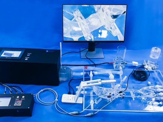
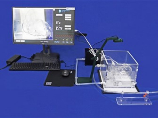
Model:Abdominal Vascular
Transarterial chemoembolization blocks the blood supply to liver tumor cells. Without affecting liver tissue, the tumor will naturally necrotize due to lack of nutrients, that is, "starve" tumor cells to death, exerting a lasting anti-tumor effect. The blood supply of the liver is different from that of other organs. 25% comes from the hepatic artery and 75% comes from the portal vein. Liver tumor tissue is almost entirely supplied by the hepatic artery. Therefore, after some hepatic arteries are embolized, normal liver tissue still has blood supply from the portal vein and will not cause necrosis.
After local anesthesia at the root of the thigh, the doctor inserts a catheter into the femoral artery under X-ray fluoroscopy, and retrogradely enters the hepatic artery, injects contrast agent, performs angiography, and determines the location, size, number, and vascular distribution of the tumor; then inserts the catheter into the artery that supplies blood to the tumor, and directly injects chemotherapy drugs or embolic agents to eliminate the tumor while blocking its blood supply, causing the tumor to be ischemic, necrotic, and atrophic. Hepatic artery chemoembolization is a minimally invasive treatment technique for liver tumors. It does not require surgery and is less toxic than systemic chemotherapy. It is a commonly used minimally invasive interventional treatment method for liver cancer patients, and has good treatment effects, which can effectively improve the quality of life of liver cancer patients.
The hepatic artery vascular model is a flat three-dimensional structure. The whole model includes the femoral artery, iliac artery, abdominal aorta and hepatic artery. There are left and right femoral artery intervention ports for doctors to learn training, such as hepatic artery vascular intervention operations.