#Product Trends
Optical and Magnetic Navigation for Tumor Ablation
Precision Targeting. Real-Time Visualization.
In radiofrequency ablation (RFA) and microwave ablation (MWA), identifying a tumor does not always ensure accurate needle placement. Preoperative imaging and intraoperative treatment often operate separately, limiting procedural precision and safety.
Key Clinical Challenges
01 Experience-Based Needle Placement
Respiratory motion and tissue deformation can shift the target after preoperative CT or MRI. Physicians must often rely on experience and repeated CT scans to adjust the needle trajectory, increasing the risk of deviation and radiation exposure.
02 Uncertain Ablation Margins
Without accurate visualization of the ablation zone, insufficient coverage may leave residual tumor, while excessive treatment can damage nearby vessels, nerves, and healthy tissue.
03 Subjective Outcome Assessment
Differences in patient position and imaging phase make pre- and postoperative scans difficult to align, reducing the accuracy and objectivity of treatment evaluation.
ARIEMEDI’s Full-Workflow Ablation Navigation Solution
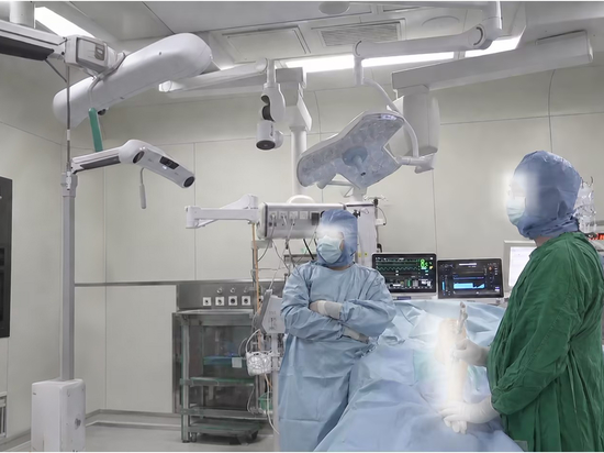
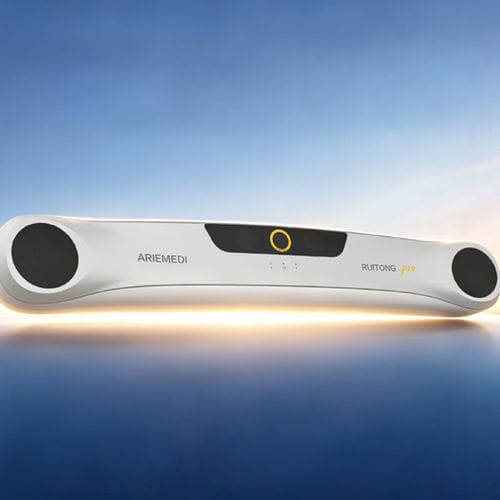
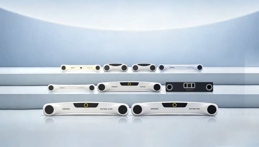
ARIEMEDI integrates preoperative planning, intraoperative navigation, and postoperative evaluation into one workflow. High-precision optical and magnetic tracking, combined with advanced medical image processing, transforms ablation from an experience-based procedure into a visualized, data-driven therapy.
01 Preoperative Planning
3D trajectory planning: Define entry points and needle paths while visualizing nearby anatomy and critical structures.
Ablation zone simulation: Predict treatment coverage based on needle type, power, and duration before puncture.
02 Intraoperative Navigation
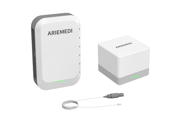
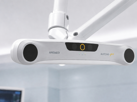
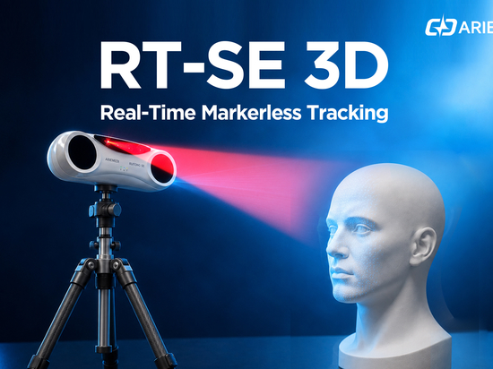
Optical and magnetic tracking: Optical tracking provides high-precision positioning and 3D surface registration, while magnetic tracking maintains reliable localization when the line of sight is obstructed.
Respiratory motion compensation: Continuously corrects target displacement caused by breathing.
Multimodal image fusion: Combines intraoperative ultrasound with preoperative CT or MRI.
Real-time needle tracking: Displays the needle’s position, angle, and depth in six degrees of freedom, helping physicians follow the planned trajectory.
03 Postoperative Evaluation
AI-assisted image registration: Aligns pre- and postoperative images using rigid and deformable registration.
Quantitative assessment: Calculates changes in target volume, residual tumor ratio, and other key metrics.
Automated reporting: Generates standardized ablation assessment reports with one click.
From image segmentation and 3D planning to real-time navigation and quantitative evaluation, ARIEMEDI enables more precise, measurable, and traceable tumor ablation.