#Product Trends
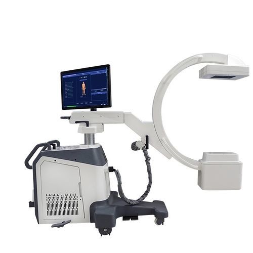
The Great C-Arm Dilemma: Image Intensifier vs. Flat Panel Detector
A Definitive Guide for Medical Practitioners
Introduction: The Evolution of Real-Time Imaging
C-arm systems revolutionized medical imaging by providing real-time X-ray visualization during surgeries, pain management procedures, and interventional diagnostics. At the heart of these systems lies a critical technological choice: traditional image intensifiers (II) or modern flat panel detectors (FPD).
1. Core Technology: How They Transform X-Rays into Images
Image Intensifiers (1950s Technology):
X-rays strike an input phosphor, converting them to electrons. These electrons are accelerated through a vacuum tube, strike an output phosphor, and create a visible light image captured by a camera. This multi-step analog process introduces geometric distortion (especially at image edges) and progressive gain degradation due to phosphor wear.
Flat Panel Detectors (1990s Technology):
X-rays strike a scintillator layer (cesium iodide or gadolinium oxysulfide), converting them to visible light. This light is immediately captured by an amorphous silicon photodetector array and converted directly into a digital signal. This streamlined process eliminates distortion and maintains consistent image quality over time.
2. Critical Comparison: Performance and Operational Impact
(table 1.jpg)
3.Clinical Applications: Matching Technology to Specialties
(table 2.jpg )
4. The Future: Where C-Arm Technology is Headed
FPDs are phasing out IIs as manufacturing costs drop and software capabilities expand. Emerging trends favor FPDs:
AI-driven image enhancement and dose management
Miniaturization for point-of-care imaging
Integration with robotic surgical platforms
Refurbished IIs will persist in price-sensitive markets, but FPDs are becoming the clinical standard.