#Industry News
The brand-new WBP equipped with a photogenetic module helps research on neurological diseases!
Successfully activates RVLM-CA neurons in conscious mice through photostimulation and records the stimulating effect on cardiovascular function
Background
Research has confirmed that activating catecholamine neurons in the ventrolateral medulla oblongata (RVLM-CA) under hypoxic conditions can enhance sympathetic nerve output, thereby increasing blood pressure (BP). To explore whether these neurons are also involved in regulating respiratory and cardiovascular variables other than blood pressure, we used the Cre-dependent carrier AAV2 EF1 α - DIO ChR2 mCherry and injected it unilaterally into the brainstem of dopamine β-hydroxylase Cre/0 mice to achieve specific expression of ChR2 mCherry by RVLM-CA neurons.
How does photostimulation of RVLM-CA neurons affect respiration and cardiopulmonary functions?
The experimental results showed that photostimulation of RVLM-CA neurons can increase the respiratory rate of anesthetized and conscious mice. In a conscious state, photostimulation mainly affects the respiratory rate, and this effect is completely suppressed under hypoxia (10% O₂) conditions. However, the effect of photostimulation remained largely unchanged under conditions of hypercapnia (3% and 6% CO₂). The related cardiovascular effects manifest as mild bradycardia and hypotension, which can be attributed to the co-activation of sympathetic and vagus nerve outputs through the analysis of selective autonomic nerve blockers. Further analysis indicates that ChR2-positive RVLM-CA neurons express VGLUT2, and their projection regions have been mapped. Its complex cardiorespiratory effects may stem from its extensive projection onto the upside spinal cord, such as the ventrolateral medulla, dorsal vagus complex, dorsal lateral pontine, and specific hypothalamic nuclei (dorsal medial, lateral, and paraventricular nuclei).
How can the small animal whole body plethysmography system assist in your research?
In the experiment, scientists used an upgraded version of a small animal whole body plethysmography system to record conscious mice's respiration changes. This upgraded version is equipped with a photogenetic model, allowing the conduct of a series of different photostimulations on animals in a conscious and freely moving state, thus achieving experimental research objectives. As shown in the above figure, the photogenetic activation of RVLM-CA neurons expressing ChR2 successfully activated the respiratory response in conscious mice. Under photostimulation of 20Hz and 1 ms, the respiratory rate significantly increases. Figure from the D experiment showed that under the conditions of 5, 10, and 20 Hz, pulse for 5 ms, conduct continuous photostimulation for 30 seconds, the respiratory rate of the experimental mice was significantly increased in direct proportion.
Solutions
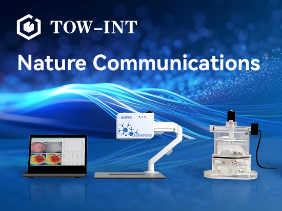
Tow-Int Tech Small Animal Whole Body Plethysmography System-Optoelectronic Version
In studies similar to those mentioned above, Tow-Int Tech can provide researchers with a complete set of experimental solutions, including the brand-new photoelectric version of the WBP-whole body plethysmography system and other expansion modules.
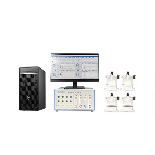
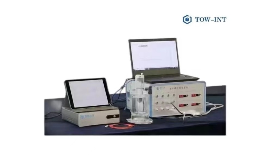
Expansion modules
The small animal whole body plethysmography system can also be combined use with small animal telemetry devices, and equipment for long-term measurement and simultaneous recording of changes in animal blood pressure and heart rate under photoelectric stimulation, to assist domestic and foreign customers in scientific research experiments.
Contact us now!
We are committed to making your research easier, more accurate, and more efficient, helping you build confidence in your data! We have provided services for a large number of customers, which helped us accumulate rich experiences in offering customized, professional solutions according to your needs.
Learn more about the whole body plethysmography system
Conclusions
In summary, this study activated RVLM-CA neurons through selective photogenetics in conscious mice, revealing important new functions of these neurons in respiratory stimulation and cardiac vagus nerve outflow control. Further research has shown that RVLM-CA neurons are likely the basis for certain acute respiratory responses induced by carotid body stimulation, making a significant contribution to the development of neurological drugs.
Reference:
Selective Optogenetic Activation of Rostral Ventrolateral Medullary Catecholaminergic Neurons Produces Cardiorespiratory Stimulation in Conscious Mice