#Industry News
Whole Body Plethysmography
Whole Body Plethysmography – in Vivo Analysis of Ventilation Function in Rodents
Background
Metabolic diseases, mainly obesity, affect over 30% of the global population and have become a major public health crisis in the past few decades (Roger et al., 2020). Meanwhile, hypertension also exists in over 30% of the global population, and there is a significant overlap between these two groups (Virani et al., 2021). Although cardiovascular disease remains the leading cause of death in the United States, the American Heart Association states that obesity and hypertension are the main factors leading to cardiovascular disease (Roger et al., 2020; Angel et al., 2020). Obesity is a complex disease characterized by energy imbalance caused by various factors such as genetics, environment, and behavior. It is estimated that human obesity results from a sustained energy imbalance of ≈ 7 kcal/d (equivalent to a typical 2000 kcal/d turnover rate of ≈ 0.35%) (Hall et al., 2011). From this, it can be inferred that such a small chronic imbalance can lead to obesity, indicating that each input and output mechanism in body efficiency plays a crucial role in controlling energy balance and the potential role of each mechanism as a cause and treatment of obesity. Therefore, careful, rigorous, reproducible, comprehensive, and synchronous research is needed on all mechanisms involved in energy flux to fully understand their impact on metabolic diseases such as obesity.
In 2019, the Wisconsin School of Medicine (MCW) established the Comprehensive Analysis Center for Rodent Metabolic Phenotypes (CRMPC) to quantitatively and comprehensively evaluate rodents’ energy and body fluid homeostasis to assist in analyzing the comprehensive mechanisms of complex metabolic diseases. A notable feature of CRMPC is its unique instrument for deep mechanical dissection of rodents’ resting metabolic rate (RMR), including anaerobic aspects that few metabolic phenotype analysis facilities can evaluate. Research has shown that the human body primarily resists weight loss through adaptation (i.e., inhibition) of RMR, and the anaerobic metabolism of gut bacteria plays a significant role in energy metabolism in humans and animals. However, this effect largely disappears during obesity. Therefore, it is necessary to develop a routine and precise workflow for evaluating gut microbiota status and develop and optimize techniques for evaluating aerobic and anaerobic metabolism in the body. This article introduces the workflow of CRMPC and MCW collaborative laboratories for comprehensive cardiac metabolic phenotype analysis of rodents and discusses the advantages and disadvantages of related equipment design for reference by researchers.
The following is a detailed introduction to unconstrained whole body plethysmography and related instruments and equipment in the main technologies.
Unconstrained Whole Body Plethysmography
Objective
Blood’s gas exchange and acid-base chemistry are intricately related to metabolic physiology. The hindbrain’s control of respiration is closely related to the hindbrain mechanism’s autonomous control of cardiovascular function. These are important bases for studying metabolic diseases such as obesity. Therefore, we need to study further rodents’ quantitative ventilation function, which can be done by unconstrained whole body plethysmography. Animals are placed in a chamber similar to a respiratory measurement chamber and then continuously exposed to an ambient air mixture with altered and known O2 and CO2 levels to evaluate changes in respiratory parameters and metabolic rates under hypoxic and hypercapnia conditions.
Introduction
Unconstrained whole-body plethysmography (WBP) is a technique that can measure respiratory rate and estimate tidal volume, allowing for the measurement and calculation of various respiratory parameters, such as minute ventilation. This method was initially established by Drorbaugh and Fenn (1955) to measure pressure deflection caused by inhalation and exhalation. In a closed chamber, there is a constant airflow (in and out) and chamber temperature. When an animal inhales, the air entering the animal is heated and humidified, causing volume expansion and increasing the pressure inside the chamber. The opposite situation occurs when exhaling. Measuring this pressure deflection can provide continuous inspiratory and expiratory waveforms for subsequent analysis and quantification of respiratory rate and tidal volume. Many teams have adopted this method to measure respiration in various species, including conscious mice (Hodges et al., 2008; Hodges and Richerson, 2008) and rats of all ages (Mouratian et al., 2012, 2017, 2019). In short, mice (all ages) and rats (<22 days old) were placed in custom 200ml or 10L plethysmography (as shown in the above figure). The gas inflow and outflow rates are balanced (150 ml/min) or slightly biased towards more outflows to prevent the accumulation of CO2. Different gases are injected into the chamber using a gas manifold connected to the gas inlet port. Use a temperature-controlled heat source to heat the chamber to ensure that the chamber temperature reaches 28-30 ° C. When measuring pressure deviation, a differential pressure sensor (Validyne) is used in units of volts produced during inhalation and exhalation while continuously measuring temperature and relative humidity (Omega). Connect the analog signal to the A/D converter and record it using a data acquisition system at a sampling rate of 200 Hz. After each experiment, a T-shaped rectal thermocouple was used to obtain the rectal temperature of each animal. Perform offline analysis of the data collected for each study. The peak value in each ventilation waveform is identified by using the “cycle measurement” option. Then, use the “height measurement” option to identify and measure the voltage deviation from the peak of each breath waveform. Use volume calibration to convert volts to volume (milliliters) and calibrate based on animal and chamber temperature, relative humidity, and ambient air pressure to estimate the tidal volume per breath. Drorbaugh and Fenn (1955) quantified respiratory rate using LabChart’s “cycle measurement” option. Minute ventilation volume (VE; ml/min) is the product of respiratory rate (F; respiration/min) and tidal volume (VT; ml/respiration). All ventilation waveforms selected for quantification are free of breathing (pauses, sighs, and smells) and motion artifacts. The measured ventilation during any state (hypoxia or hypercapnia) is divided by indoor air respiration and multiplied by 100% to evaluate the sensitivity of the respiratory system to oxygen and carbon dioxide.
Advantages and Disadvantages
The whole body plethysmography can provide a minimally invasive respiration assessment without the need for anesthesia, and this method can be reused with other methods. For example, the system is also multiplexed with antennas for wireless telemetry of blood pressure or other endpoints and for manipulating stimulators for optogenetics. The disadvantage of this method is that it cannot evaluate lung resistance and compliance.
Solutions
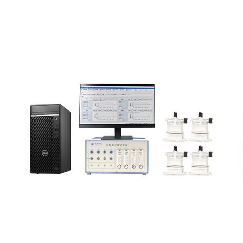
Tow-Int Tech-Whole Body Plethysmography System (WBP)
The whole body plethysmography (WBP) system developed by Tow-Int Tech can measure of conscious and free-moving animals, such as respiratory rate, tidal volume, and airway hyperresponsiveness (AHR) testing. During the testing process, animals can be conscious and free, avoiding the impact of traumatic tracheostomy and anesthesia, making the experimental process more convenient. It is used to study the responsiveness of respiratory system model animals to drugs and the pharmacology and toxicology of respiratory drugs. It is particularly suitable for rapid screening experiments of large-scale animals, long-term tracking studies, and repetitive screening.
Features-Tow-Int Tech WBP
Applicable animals: mice, young mice, rats, guinerespiratory parametersa pigs, rabbits, dogs, cats, miniature pigs, monkeys, and other animals
Equipped with a feeding and water inlet device to facilitate long-term experiments
Measurement channels: 1-64 channels
Automatic bias control function
Configurable high-frequency oscillation atomization drug delivery system
Special noise reduction structure design can effectively reduce interference caused by environmental changes
Can perform noise reduction in the bias flow meter, improving the signal-to-noise ratio of the signal and reducing system noise
Support external nitrogen or other gases to complete hypoxia experiments
Equipped with analysis software, data can be saved in Excel or txt format
The software can automatically switch up to four different gas channels and can be operated through an external controller
*Various professional solutions can be customized according to customer experimental needs
Contact us now!
We are committed to making your research easier, more accurate, and more efficient and helping you build confidence in your data! We have provided services for a large number of customers, giving us rich experiences in offering customized, professional solutions according to your needs.
Learn more about the whole body plethysmography system