#Industry News
Application of Whole Body Plethysmography System (WBP)
in the Study of Pulmonary Fibrosis
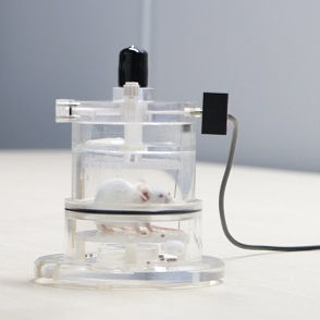
Whole Body Plethysmography System-WBP
The whole body plethysmography system can measure the respiratory parameters of conscious and freely moving animals, such as respiratory rate, tidal volume, airway hyperresponsiveness (AHR), etc. During the test, the animal can be conscious and unrestrained, avoiding the impact of traumatic tracheotomy and anesthesia, making the experimental process simpler. It is used to study the responsiveness of respiratory model animals to drugs and the pharmacology and toxicology of respiratory drugs. It is particularly suitable for large-scale rapid initial screening tests of animals, long-term follow-up studies, and repeated screening. Whole body plethysmography systems are also used in the study of pulmonary fibrosis.
Background
At present, the incidence and mortality of pulmonary fibrosis are increasing year by year. At the same time, there is still a lack of effective treatment methods in clinical practice. Studies have shown that mesenchymal stem cells can treat lung inflammation and alleviate the progression of pulmonary fibrosis through multiple mechanisms, including paracrine effects, mitochondrial transfer, and induction of macrophage polarization. However, stem cell therapy still faces problems such as difficulty monitoring stem cells after transplantation, short lung retention time, and low survival rate.
A New Multifunctional Probe Assisting in Stem Cell Therapy
In response to the above problems, the team of researcher Chen Hangrong and associate researcher Ma Ming from the Shanghai Institute of Ceramics, Chinese Academy of Sciences, used the "metalloporphyrin-induced multiscale self-assembly" strategy to construct a new multifunctional probe assisting in stem cell therapy. The composite probe comprises three functional components with clinical application potential: cobalt chloride protoporphyrin (CoPP), gold nanoparticles, and superparamagnetic Fe3O4 nanoparticles. It uses copper-free click chemistry to achieve efficient bioorthogonal labeling of stem cells. This self-assembled nanoprobe does not require additional carriers and surfactants and can effectively exert the corresponding functions of each component at a lower dose. Specifically, the two functions of CT contrast imaging and magnetic manipulation of the nanoprobe respectively solve the problems of difficult monitoring and short retention time of stem cells in the lungs; the probe effectively improves the survival rate and repair effect of stem cells by slowly releasing the antioxidant drug CoPP in the stem cell microenvironment. By further constructing a small animal model of pulmonary fibrosis, it was confirmed that compared with the single stem cell treatment group, this multifunctional self-assembled nanoprobe-mediated stem cell therapy can significantly alleviate fibrosis symptoms and promote lung function recovery and has good clinical application potential.
Schematic diagram of the preparation of the multifunctional probe; (b) Schematic diagram of the probe used to label stem cells and treat pulmonary fibrosis; (c) TEM image of the bowl-shaped multifunctional probe; (d) High-angle annular dark field image and element distribution image; (e) Microscopic image of stem cells labeled by the multifunctional probe, the red arrow indicates the probe particles inside the cell.
Bleomycin-induced Pulmonary Fibrosis Model
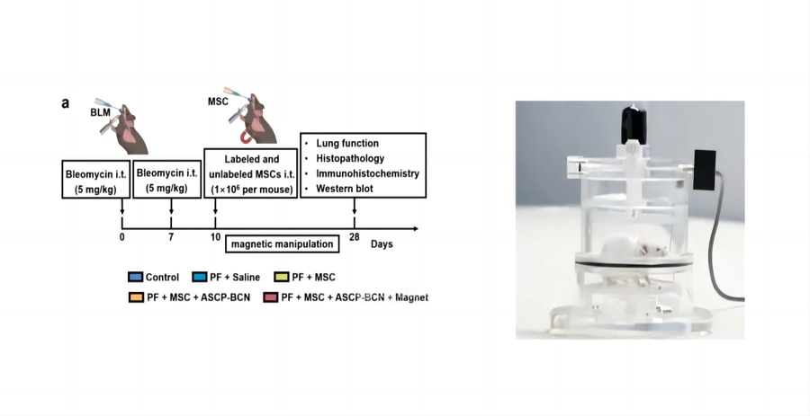
36 male C57BL/6 mice were acclimated for 1 week and randomly divided into 6 groups: (1) control, (2) PF, (3) PF + saline, (4) PF + MSC, (5) PF + ASCP-BCN labeled MSC, (6) PF + ASCP-BCN labeled MSC + magnet (n = 6). Pulmonary fibrosis was induced with bleomycin (BLM). Briefly, mice were anesthetized, and BLM (5 mg/kg) was dissolved in 100 μL sterile saline and slowly injected into the tracheal lumen through an endotracheal cannula. Mice in the saline group were given only 100 μL sterile saline. The above steps were repeated on the 7th day after administration. Unlabeled MSCs and labeled MSCs (1×10⁶ cells per mouse) were placed in 100 μL saline and transplanted intratracheally into the mouse lungs on the 10th day after the first injection of BLM. In the PF + ASCP-BCN labeled MSC + magnet group, a NdFeB magnet was attached to the chest of each mouse. Mice were executed on day 28 after the first BLM injection, and lung tissues were obtained.
Bone Marrow Mesenchymal Stem Cell Transplantation
On day 28, after the first BLM injection, the mice were weighed and placed in a WBP whole body plethysmography system to record their lung function data. The lungs of the mice were then collected for further identification. One lung lobe of each mouse was taken, and the blood on its surface was gently wiped. The lung tissue was weighed and baked at 60°C for 72 h. The dry lung tissue was weighed again, and the wet-to-dry ratio of the lung was determined. The HYP concentration in the lung tissue was detected using the HYP detection kit.
Therapeutic Effects
Therapeutic effects of ASCP-BCN-labeled MSCs and Magnetic Manipulation on PF
(a) Experimental scheme of MSC treatment of bleomycin-induced PF mouse model. (b-e) Lung function of mice without treatment (control) and bone marrow-induced PF mice model with different treatments of (saline, MSCs, ASCP-BCN-labeled MSCs, and ASCP-BCN-labeled MSCs + magnetic manipulation) for 28 days, then complete the first injection of bone marrow:
(b) Breaths per minute, (c) 50% vital capacity of expiratory flow, (d) peak inspiratory flow, (e) peak expiratory flow.
(f) Content of HYP in the lung.
(g) Wet-to-dry ratio of the lungs of each group.
(h) Representative H&E staining images, Masson trichrome staining images, and α-SMA immunohistochemical staining images of lung sections of each group.
(i) The severity of PF was evaluated by the Ashcroft score corresponding to H&E staining.
(j) Masson trichrome staining was used to determine the percentage of lung collagen deposition area.
(k) Immunohistochemical staining shows the percentage of α-SMA positive cells.
(l) Immunoblot analysis of α-SMA and TGF-β in lung tissues.
(m) Relative expression levels of the corresponding α-SMA and TGF-β proteins.
The therapeutic effect of BMSCs on PF
To evaluate the feasibility of ASCP-BCN-labeled BMSCs in the treatment of PF, a bleomycin (BLM)-induced mouse PF model was used. BLM intervention led to a significant decrease in lung function in mice, as manifested by an increase in minute volume (MV), expiratory flow at 50% vital capacity (EF50), peak inspiratory flow (PIF), and peak expiratory flow (PEF). BMSC transplantation slightly reduced the above lung function parameters. ASCP-BCN-labeled BMSC transplantation and magnetic attraction further alleviated the above lung function parameters, indicating that it plays an important role in promoting the therapeutic effect of BMSCs. In particular, mice's respiratory rate and peak inspiratory flow in the ASCP-BCN-labeled MSCs + magnetic manipulation transplantation group almost reached those of the control group. More importantly, labeled MSC transplantation and magnetic manipulation not only reduced the hydroxyproline (HYP) concentration in fibrotic lungs but also reduced the lung wet-to-dry ratio caused by inflammation.
Related Research
The results were recently published in the journal Bioactive Materials (2023, 21, 129) under "Cobalt protoporphyrin-induced nano-self-assembly for CT imaging, magnetic-guidance, and antioxidative protection of stem cells in pulmonary fibrosis treatment".
Related Links
https://www.sciencedirect.com/science/article/pii/S2452199X22003383
https://www.sciencedirect.com/science/article/pii/S1748013220300669
Contact us now!
We are committed to making your research easier, more accurate, and more efficient and helping you build confidence in your data! We have provided services for a large number of customers, giving us rich experiences in offering customized, professional solutions according to your needs.