#Product Trends
ERCP (Endoscopic Retrograde Cholangiopancreatography)
ERCP (Endoscopic Retrograde Cholangiopancreatography)
ERCP stands for endoscopic retrograde cholangiopancreatography. It’s a type of imaging test that allows healthcare providers to look inside your bile ducts and pancreatic ducts.During ERCP, healthcare providers use an endoscope and X-rays to observe injectable dye traveling through your pancreatic and bile ducts.
Why would I need an ERCP?
Your provider might suggest an ERCP if you have symptoms that suggest a problem in your biliary system, such as:
*Unexplained upper abdominal pain or biliary colic.
*Signs of stalled bile flow or bile leakage, like jaundice.
The ERCP procedure can help diagnose and treat common issues affecting your biliary ducts, such as:
*Inflammation (cholangitis) and possible infection.
*Narrowing caused by scar tissue (biliary stricture).
*Gallstones in your common bile duct.
*Gallstone pancreatitis.
*Tumors or cancer in your bile ducts.
*Bile duct leaks or other injuries.
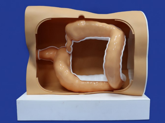
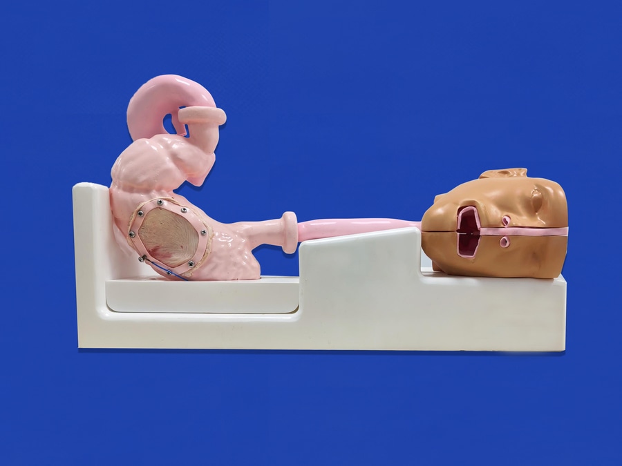
Trandomed’s ERCP model is constructed using CT data from the adult male digestive system and relevant anatomical literature. It is composed of soft silicone and produced through 3D printing technology. The model include the head, esophagus, stomach, duodenum and etc, and it could be designed and replaced in sections. The main usage of the model is for training in upper gastrointestinal endoscopy, and other endoscopic examinations. Besides that, the model can be used for ERCP path operation, like duodenal papilla insertion, bile drainage, biliary stone extraction and other operations.
What is adrenal venous sampling?
Adrenal venous sampling is a procedure which involves inserting a small plastic tube into the veins from the adrenal glands and taking a small sample of blood. This sample is then sent to the laboratory for testing. Normally veins do not show up on an ordinary x-ray so a special dye, called contrast medium, is injected into the veins through a fine plastic tube called a catheter to make them visible. If unilateral, the adrenal gland can be removed; thus curing secondary hypertension in 50 to 80% of the cases that are caused by aldosterone-producing adenoma while the remaining cases show improvement in hypertension treatment 1. If bilateral, the hypertension is better controlled medically with aldosterone antagonists.
The AVS procedure is as follows:
1)Preoperative preparation: Patients undergo preoperative cross-sectional imaging studies (such as adrenal CT) to determine the location, size, and morphology of bilateral adrenal lesions and the anatomical position of the bilateral adrenal veins. To minimize interference with blood sampling results, medications that interfere with the renin-angiotensin-aldosterone system should be discontinued preoperatively;
2)Surgical approach and catheter selection;
3)Adrenal vein catheterization, angiography, and blood sampling;
4)Result analysis: SI index, LI index;
In the analysis of AV results, the SI, renal, and LI numbers need to be counted on the fingers. A thorough understanding of the location of the vein opening and angiographic morphology is crucial to improving the success rate of blood collection.
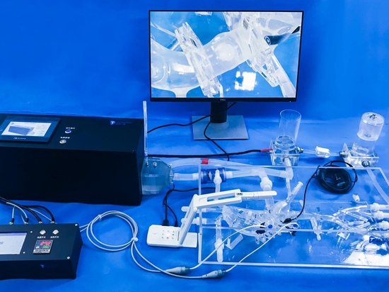
The AVS training simulator is a clinical training simulator developed for adrenal venous sampling (AVS). Using a realistic silicone vein model and simulated DSA vascular imaging equipment, it guides trainees through catheterization and adrenal venous angiographic morphology recognition, improving learning and training efficiency.
The venous vascular model, designed based on CT data and anatomical literature, includes multiple adrenal vein openings and interfering veins such as the accessory hepatic vein, providing a more realistic vascular structure for operator training. It also provides simulated DSA vascular imaging equipment. Through acquisition devices and data processing, the position of the operating instrument relative to the spine can be observed in real time under simulated DSA images, enabling observation of simulated DSA imaging in a radiation-free environment and judgment based on the imaging results. It is compatible with basic visualization training and advanced DSA-simulated catheter intubation and angiography training.