#Product Trends
Ophthalmic Artery Aneurysm: A Hidden Danger Near the Eye
Ophthalmic Artery Aneurysm: A Hidden Danger Near the Eye
The ophthalmic artery aneurysm is a rare but potentially dangerous vascular condition occurring near the junction where the ophthalmic artery branches from the internal carotid artery (ICA). Because of its deep location and proximity to critical structures like the optic nerve, early diagnosis and precise intervention are essential.
1. What Is an Ophthalmic Artery Aneurysm?
An aneurysm is a bulging or ballooning of a blood vessel wall caused by weakening of the vessel tissue. In the case of an ophthalmic artery aneurysm, this abnormal dilation develops at or near the origin of the ophthalmic artery, which supplies blood to the eye and surrounding tissues. If the aneurysm ruptures, it can lead to subarachnoid hemorrhage, a life-threatening form of brain bleeding.
2. Symptoms and Clinical Manifestations
Because of its location, the most common symptoms are visual disturbances, such as:
• Blurred vision or double vision
• Loss of part of the visual field
• Pupil abnormalities or drooping eyelid
Some patients remain asymptomatic until the aneurysm grows large enough to compress the optic nerve or ruptures suddenly, causing severe headache and neurological symptoms.
3. Diagnosis and Imaging
CT angiography (CTA), MR angiography (MRA), and especially digital subtraction angiography (DSA) are key imaging tools used to detect ophthalmic aneurysms and to guide treatment. Advanced 3D reconstruction technologies now allow medical professionals to visualize the aneurysm’s size, neck morphology, and spatial relationship to surrounding arteries.
4. Treatment and Intervention
Treatment typically involves microsurgical clipping or endovascular coiling/stenting, depending on the aneurysm’s size and shape. Endovascular techniques are increasingly preferred for their minimal invasiveness and high precision, but they require accurate preclinical testing and operator training.
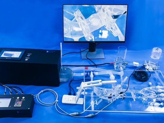
5. How the Simulation Model Helps
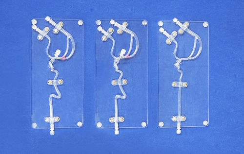
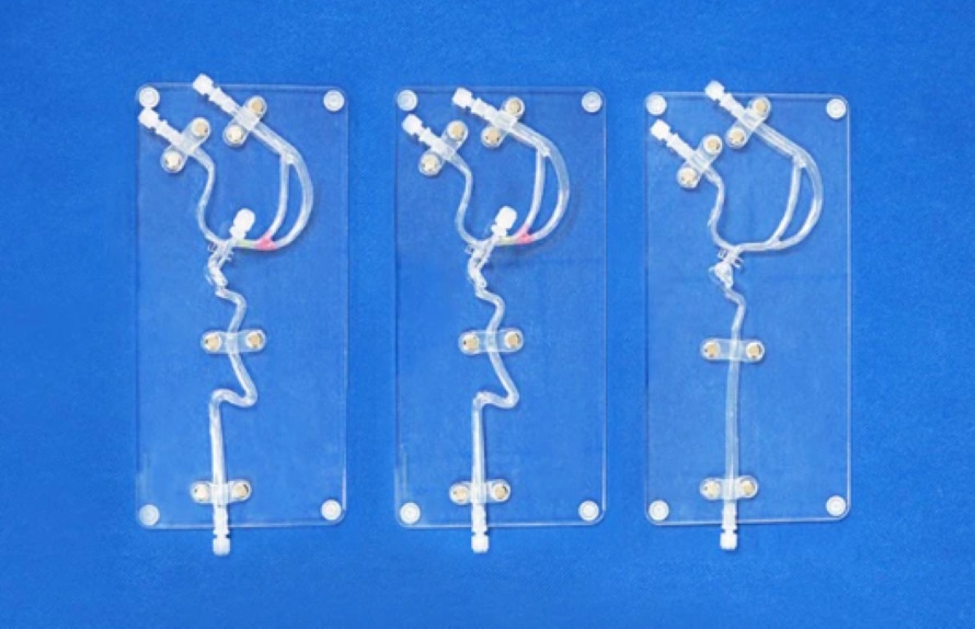
The Internal Carotid and MCA Model is designed for realistic simulation of ophthalmic artery aneurysm interventions. It replicates the 1:1 anatomical structure of the ICA and MCA based on CT-derived data. The model allows users to:
• Evaluate catheter and guidewire trackability through tortuous arteries
• Practice aneurysm coiling and stent-assisted embolization
• Customize aneurysm number, size, and position to simulate different clinical scenarios
• Study hemodynamic flow and deployment precision under realistic vascular conditions
Such models provide a safe and repeatable environment for physicians and device developers to test new techniques and optimize performance before clinical application.
In summary, ophthalmic artery aneurysms may be small and hidden, but their consequences can be severe. With the aid of advanced anatomical simulation models, clinicians and researchers can better understand this delicate region, refine interventional techniques, and ultimately improve patient safety and treatment outcomes.