#Product Trends
When Back Pain Feels Like a Knife—Could It Be Kidney Stones?
When Back Pain Feels Like a Knife—Could It Be Kidney Stones?
Kidney stones are more common than many think. Our kidneys act like two sophisticated filters, processing around 180 liters of blood each day and producing 1–2 liters of urine. During this process, certain minerals may crystallize—just like limescale forming in a kettle—gradually building up to form stones.
Common Types of Kidney Stones
1.Calcium stones (≈80%) – often calcium oxalate or calcium phosphate
2.Uric acid stones – frequently seen in people with gout
3.Infection-related stones – associated with urinary tract infections
4.Cystine stones – rare and linked to genetic conditions
Many stones remain silent for a long time. However, once they move and become stuck in the ureter (the thin tube carrying urine to the bladder), they cause renal colic—one of the most severe types of pain known in medicine.
Why Is the Pain So Intense?
The ureter is extremely narrow—only 2–3 mm wide. When a stone blocks it:
1.Urine backs up, increasing pressure inside the kidney
2.The ureter contracts forcefully to push the stone downward
3.Pain signals spread along nerves to the abdomen, groin, or genital area
Symptoms often include:
1.Sudden, severe one-sided back or flank pain
2.Pain that comes in waves, becoming increasingly intense
3.Nausea, sweating, or vomiting
4.Possible blood in the urine
What Increases the Risk of Kidney Stones?
1.Drinking too little water
2.High-salt or high-protein diets
3.Frequent intake of high-oxalate foods (such as spinach and nuts)
4.Sedentary lifestyle
5.Family history, gout, or certain medications
How Are Kidney Stones Diagnosed?
1.Urinalysis to check for blood or infection
2.Ultrasound for safe, radiation-free imaging
3.CT scan, which provides the most accurate diagnosis
Treatment Options (Rewritten Paragraph)
The choice of treatment depends largely on the size and location of the kidney stone. For stones smaller than 5 mm, conservative management is often recommended, which includes drinking plenty of water (usually 2–3 liters per day), taking pain relief medications, and engaging in moderate physical activity to encourage the stone to pass naturally. When the stone is approximately 5–10 mm, extracorporeal shock wave lithotripsy (ESWL) may be used to break the stone into smaller pieces that can be passed more easily. For larger stones or those causing severe blockage, minimally invasive surgical procedures such as ureteroscopic stone removal or percutaneous nephrolithotomy may be necessary to safely extract the stone and restore normal urinary flow.
How to Prevent Recurrence
1.Drink enough water to produce ≥2 liters of urine daily
2.Limit salt intake (<5 g/day)
3.Consume a normal, not excessive, amount of calcium
4.Reduce intake of high-oxalate and high-purine foods
5.Exercise regularly
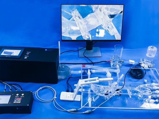
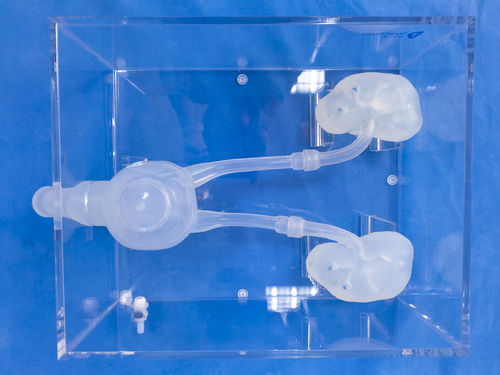
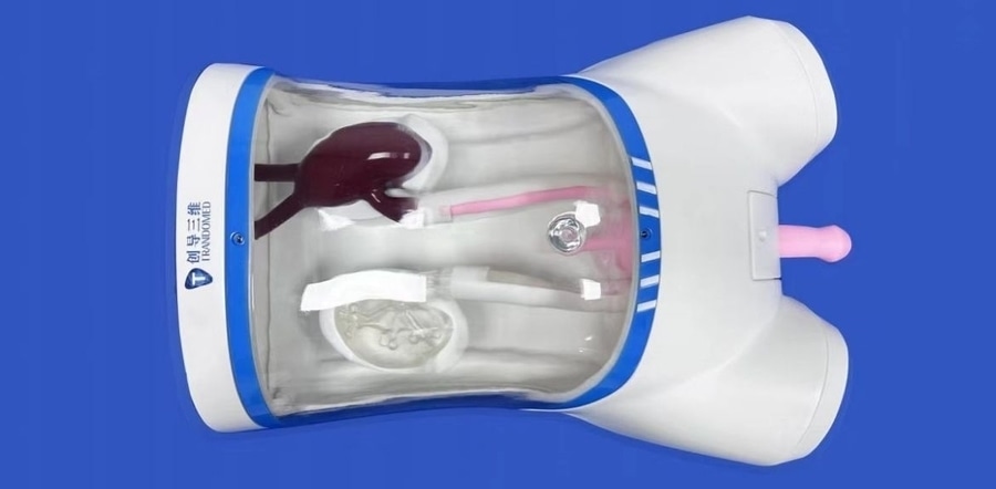
Introducing Our 3D-Printed Ureteroscopy Training Model
To support medical education and improve surgical proficiency, we offer a high-fidelity 3D-printed ureteroscopy training model designed to realistically simulate urinary tract anatomy and stone removal procedures.