#Product Trends
Understanding Patent Ductus Arteriosus (PDA)
Understanding Patent Ductus Arteriosus (PDA)
1. What is the Ductus Arteriosus?
The ductus arteriosus is a normal fetal blood vessel that connects the pulmonary artery to the aorta. This vessel allows oxygen-poor blood to bypass the non-functioning fetal lungs and flow directly into the aorta, helping to distribute blood throughout the fetus via the placenta.
Normally, one end of the ductus originates from the left pulmonary artery, and the other connects to the aortic arch, usually near the origin of the left subclavian artery.
In some rare cases, the ductus may arise from the right pulmonary artery or connect to other arteries, such as the innominate artery.
Even more rarely, double ductus arteriosus may exist, particularly in complex congenital heart conditions where the pulmonary arteries are disconnected.
2. Fetal Circulation and Hemodynamics
During fetal life:
The ductus arteriosus is essential for normal circulation because the fetal lungs are not inflated and have high resistance to blood flow.
Most blood pumped by the right ventricle bypasses the lungs through the ductus arteriosus and flows into the aorta, eventually reaching the placenta for gas exchange with the mother.
3. Closure of the Ductus Arteriosus
After birth, the ductus arteriosus is supposed to close in two phases:
1. Functional (Physiological) Closure
After the first breath, the lungs expand, pulmonary resistance drops, and blood begins to flow into the lungs instead of through the ductus.
Rising oxygen levels in the blood trigger smooth muscle contraction in the ductal wall, reducing the blood flow through the ductus.
This usually occurs within 10–15 hours after birth, although the ductus may reopen within the first 7–8 days if full closure hasn't occurred.
2. Anatomical Closure
Over the next few weeks, the ductus undergoes fibrous thickening and becomes the ligamentum arteriosum.
In 88% of infants, complete closure occurs within 8 weeks.
If the ductus remains open after 6 to 12 months, it is called Patent Ductus Arteriosus (PDA) and is unlikely to close without intervention.
4. Types and Shapes of PDA
PDA can vary in shape, size, and length. Common classifications include:
1.Tubular (Cylindrical) – Long and evenly shaped; most common (over 80% of cases).
2.Funnel-shaped – Wide at the aortic end, narrow at the pulmonary end.
3.Window-type – Very short duct forming a direct opening between the aorta and pulmonary artery.
4.Aneurysmal – Enlarged in the middle, sometimes with thrombosis.
5.Hourglass (Dumbbell-shaped) – Narrow in the middle, wider at both ends.
5. How is PDA Diagnosed?
Heart Murmur: A continuous murmur heard with a stethoscope is often the first sign.
Chest X-ray: May show enlarged heart and increased pulmonary blood flow.
Echocardiogram (Ultrasound): The most reliable diagnostic tool. It can visualize the PDA, measure its size, and assess the heart’s function.
Even in older children with no visible X-ray changes, echocardiography can detect PDA.
6. How is PDA Treated?
Observation and Medication (in newborns):
In the neonatal period, PDA may close spontaneously.
If symptoms like heart failure are mild and medication is effective, doctors may wait and observe.
Medications like indomethacin or ibuprofen can help close the PDA by causing the ductal smooth muscle to contract.
However, these drugs can have side effects like kidney issues or bleeding, so blood tests are required before use.
Surgical or Interventional Closure (after neonatal period):
If PDA does not close on its own, or if complications arise, closure is necessary to prevent infective endocarditis and pulmonary hypertension.
Catheter-based closure involves inserting a device (e.g. coil or plug) through a vein to block the PDA.
If the PDA is large or has unusual anatomy, surgical ligation through a small chest incision may be needed.
Both catheter and surgical treatments are generally safe, and risks are more related to underlying conditions than the procedure itself.
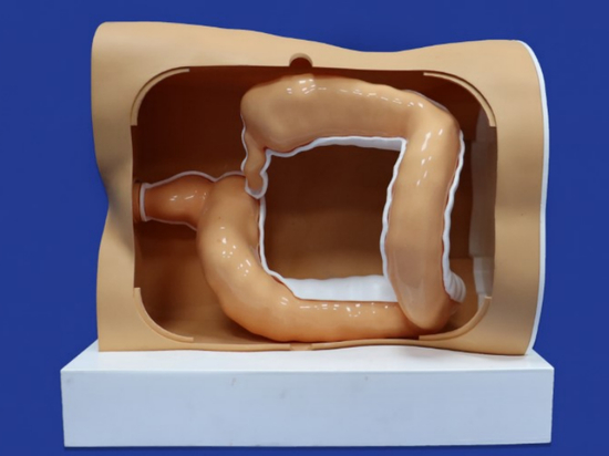
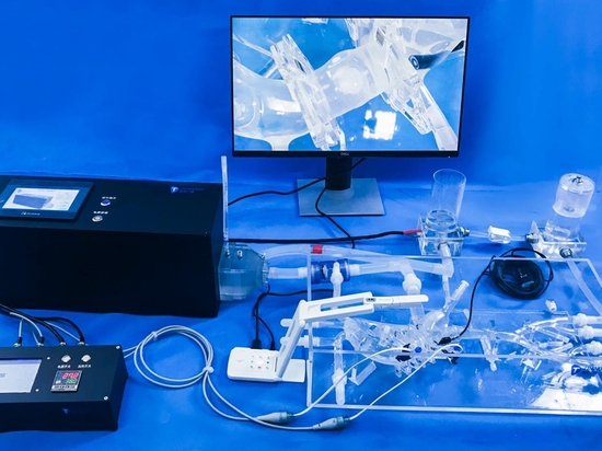
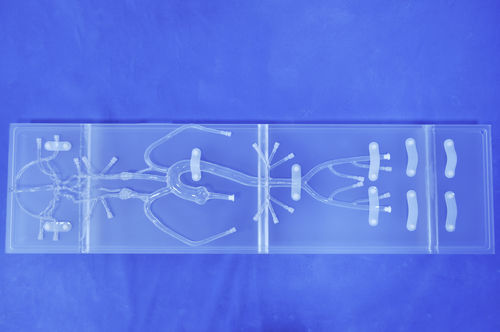
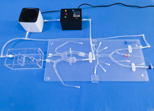
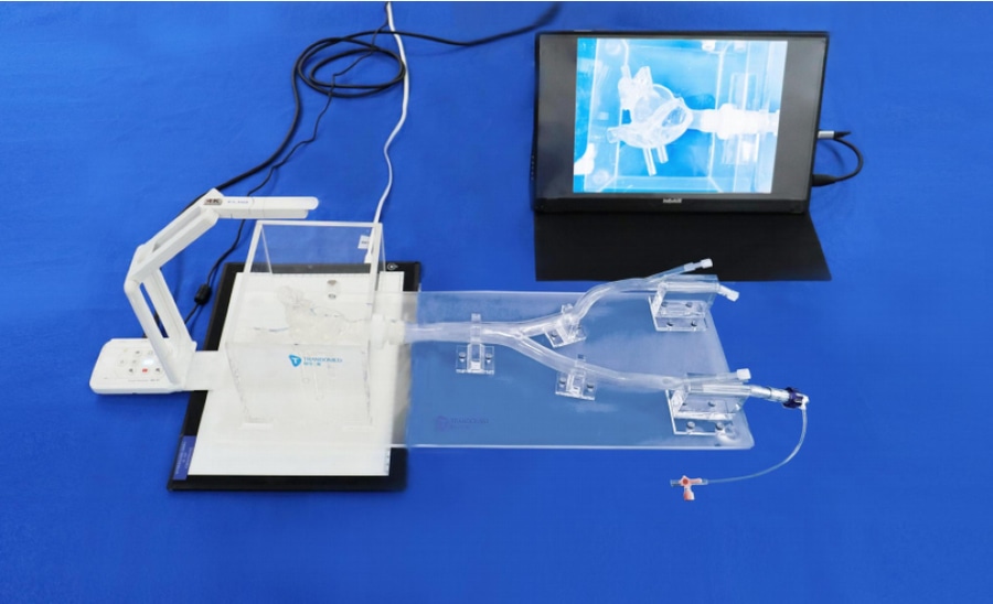
In our congenital model, we can customize PDA for you to simulate the procedure.