#Industry News
Neurovascular interventional training models are changing the growth path of young doctors
Neurovascular interventional training models are changing the growth path of young doctors
The Steep Learning Curve and High Risks of Neurointervention Training
Neurointerventional surgery, often described as "dancing on millimeter-sized blood vessels," presents a level of difficulty and risk far exceeding that of ordinary clinical surgery. Intracranial nerves and blood vessels are as thin as a hair, only a few millimeters in diameter, and are intricately intertwined with numerous branches. Their walls are extremely fragile; any minor operational error-whether it's excessive guidewire pushing force, incorrect path selection, or inaccurate microcatheter placement-can lead to serious complications such as vessel rupture and embolism, even endangering the patient's life. For resident physicians and young doctors, the path to learning neurointervention using the Neuro Test Model has always been fraught with peril, with the biggest challenge being the lack of safe, repeatable, and highly realistic practice environments. In addition, traditional residency training faces a core pain point: high pressure and high teaching risk for instructors. Instructors must ensure surgical safety while also teaching young doctors, often finding themselves in a dilemma of "not daring to let go and being unable to teach effectively."v
High-Fidelity Modeling and Zero-Risk Skill Acquisition for Young Physicians
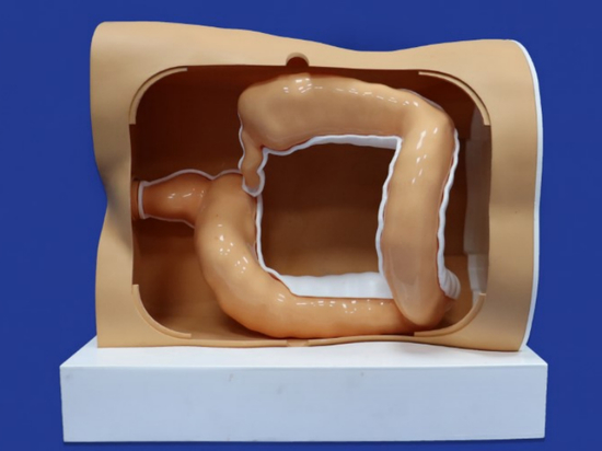
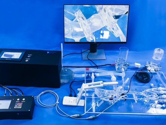
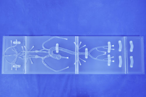
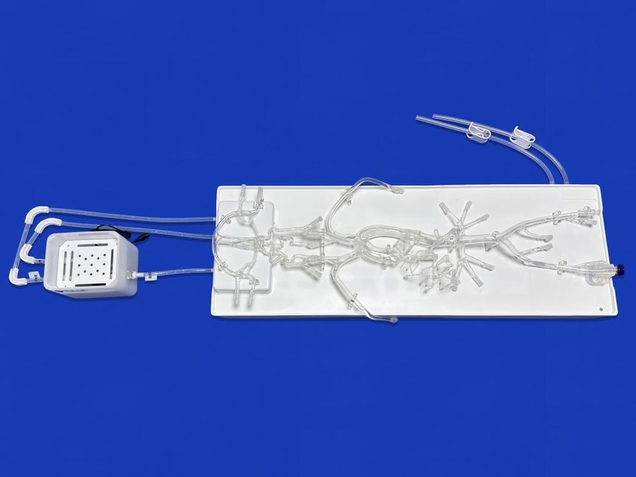
The emergence of neurovascular interventional training models precisely addresses these core contradictions in neurovascular interventional residency training, fundamentally changing the growth path of young physicians. A high-quality neurovascular interventional training model, reconstructed from real human intracranial CT and MRI image data, accurately replicates the anatomical structure of intracranial blood vessels in a 1:1 ratio. From the Circle of Willis, the middle cerebral artery, and the anterior cerebral artery to the smallest terminal branches, every tortuous angle, vessel diameter, and wall thickness is highly consistent with the real human body, even reproducing the subtle texture and elasticity of blood vessels. More importantly, the model can accurately simulate the tactile feedback during clinical practice. The resistance when pushing the guidewire, the flexibility when turning the microcatheter, and the elastic feedback of the vessel wall are all completely synchronized with real surgery, allowing young physicians to repeatedly practice the entire process of guidewire manipulation, microcatheter placement, path selection, angiography interpretation, and simulated lesion treatment in a zero-risk environment.
Standardized Teaching Platforms and Enhanced Instructor Efficiency
For department directors and supervising physicians, neurovascular interventional training models are also a powerful tool for improving residency training efficiency and standardizing teaching procedures. Through a unified simulation training platform, instructors can develop standardized teaching plans, provide precise guidance and assessment for young doctors' procedures, and promptly correct any non-standard operating techniques, ensuring that every resident physician masters core skills. Simultaneously, model training reduces teaching risks, avoiding medical hazards caused by errors made by young doctors, making mentorship more efficient, safer, and more aligned with clinical needs.