#Product Trends
Learn what microfracture surgery is, how it is performed arthroscopically, which knee cartilage defects may be considered for the procedure, and how microfracture instruments are used to stimulate a healing response from the subchondral bone.
Learn what microfracture surgery is, how it is performed arthroscopically, which knee cartilage defects may be considered for the procedure, and how microfracture instruments are used to stimulate a healing response from the subchondral bone.
Articular cartilage covers the ends of bones inside the knee and helps create a smooth, low-friction surface for joint movement.
When a focal cartilage defect develops, the damaged area may not heal effectively on its own because articular cartilage has limited intrinsic regenerative capacity.
One surgical option used for selected focal cartilage defects is microfracture surgery.
Microfracture is a marrow stimulation technique performed arthroscopically. Small perforations are created in the subchondral bone beneath the cartilage defect, allowing blood and bone marrow elements to enter the treated area.
The goal is to stimulate formation of a repair tissue over the cartilage defect.
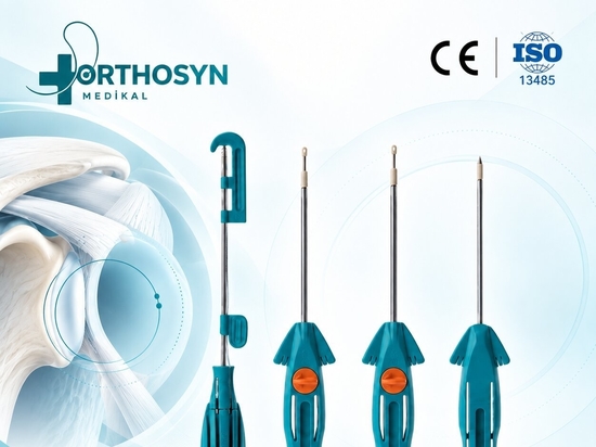
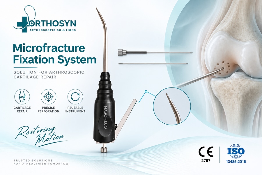
Orthosyn Medikal provides microfracture instrumentation designed for arthroscopic access and controlled subchondral perforation during selected cartilage repair procedures.
What Is Articular Cartilage?
Articular cartilage is the smooth tissue covering the ends of bones inside synovial joints.
In the knee, it covers important surfaces of the:
Femur
Tibia
Patella
Healthy cartilage helps provide:
Low-friction movement
Load distribution
Shock absorption
Protection of the underlying bone
Because cartilage contains very few blood vessels, its ability to heal after injury is limited.
This is one reason focal cartilage lesions can remain symptomatic.
What Is a Knee Cartilage Defect?
A cartilage defect is a localized area where articular cartilage has been damaged or lost.
Cartilage defects may occur because of:
Sports injury
Direct trauma
Repetitive mechanical loading
Osteochondral injury
Patellar instability
Associated ligament injury
Previous surgery
The defect may vary in:
Size
Depth
Location
Shape
Condition of the surrounding cartilage
Condition of the underlying bone
These characteristics influence treatment selection.
What Is Microfracture Surgery?
Microfracture is an arthroscopic marrow stimulation procedure used for selected focal cartilage defects.
The damaged cartilage is first evaluated and prepared.
After unstable cartilage tissue is removed, small holes are created in the exposed subchondral bone.
These perforations allow blood and marrow-derived cells to reach the defect.
The biological concept is:
Cartilage Defect → Subchondral Perforation → Marrow Access → Clot Formation → Repair Tissue
The resulting repair tissue is intended to cover the defect and improve the local joint surface.
Why Is It Called “Microfracture”?
The name comes from the creation of multiple small perforations in the subchondral bone.
These perforations are not the same as a traumatic fracture.
Instead, they are deliberately created in a controlled surgical pattern to access the underlying marrow environment.
The term microfracture therefore refers to a marrow stimulation technique rather than an accidental bone fracture.
How Is Microfracture Performed?
Although surgical techniques can vary, the general procedure usually includes several steps.
1. Arthroscopic Evaluation
The surgeon first inspects the knee arthroscopically.
The cartilage defect is evaluated for:
Location
Size
Depth
Stability of surrounding cartilage
Associated pathology
2. Defect Preparation
Loose or unstable cartilage is removed.
The edges of the defect are prepared so that stable cartilage surrounds the treated area.
The calcified cartilage layer may also be carefully prepared depending on the technique.
3. Subchondral Perforation
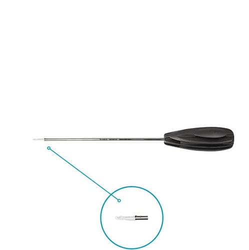
A microfracture awl or similar instrument is used to create small perforations in the subchondral bone.
4. Confirmation of Marrow Access
After perforation, blood or marrow elements may be observed entering the defect.
This indicates communication with the underlying marrow space.
5. Rehabilitation
Postoperative rehabilitation is an important component of the overall treatment strategy.
The exact protocol depends on:
Defect location
Defect size
Weight-bearing surface
Associated procedures
Surgeon preference
What Happens After the Bone Is Perforated?
When the subchondral bone is perforated, blood and marrow-derived components enter the cartilage defect.
These can form a clot over the treated area.
The clot contains biological elements involved in tissue repair.
Over time, this may develop into repair tissue that fills part or all of the defect.
However, the tissue formed after microfracture is not identical to native hyaline articular cartilage.
It is commonly described as fibrocartilaginous repair tissue.
This distinction is important when discussing the biological objective of microfracture surgery.
Is Microfracture the Same as Cartilage Regeneration?
Not exactly.
Microfracture does not simply recreate normal native articular cartilage.
The procedure stimulates a marrow-derived healing response intended to produce repair tissue within the defect.
The repair tissue may contain fibrocartilage characteristics rather than the same structural composition as normal hyaline cartilage.
For this reason, microfracture is more accurately described as a cartilage repair or marrow stimulation technique rather than complete cartilage regeneration.
Which Knee Cartilage Defects May Be Considered for Microfracture?
Microfracture may be considered for selected focal, full-thickness cartilage lesions.
Patient selection depends on multiple factors, including:
Defect size
Defect location
Depth
Patient age
Activity level
Body weight
Alignment
Meniscal condition
Ligament stability
Previous surgery
Condition of surrounding cartilage
Microfracture is not automatically appropriate for every cartilage defect.
The complete condition of the knee must be evaluated.
Why Does Defect Size Matter?
Defect size can influence treatment strategy.
Microfracture is generally discussed in relation to focal cartilage defects, rather than widespread loss of cartilage throughout the joint.
A larger defect creates a greater area that must be filled by repair tissue.
The size of the lesion may therefore influence:
Biological repair potential
Mechanical environment
Rehabilitation
Expected durability
Choice between cartilage repair techniques
Treatment decisions should not be based on size alone, but it is an important factor.
Why Does Defect Location Matter?
Different regions of the knee experience different mechanical loads.
Cartilage defects may occur on:
Medial femoral condyle
Lateral femoral condyle
Trochlea
Patella
Tibial plateau
A defect on a major weight-bearing surface experiences different loading from one in another part of the joint.
Location can therefore influence:
Surgical access
Instrument angle
Rehabilitation
Weight-bearing restrictions
Treatment selection
What Is Subchondral Bone?
The subchondral bone is the layer of bone directly beneath the articular cartilage.
It provides structural support to the cartilage surface.
In microfracture surgery, the surgeon intentionally penetrates this layer to gain access to the marrow space beneath it.
The objective is not to remove large amounts of bone.
Instead, controlled small perforations are created to stimulate a biological healing response.
Why Is Hole Placement Important?
The microfracture perforations should be distributed across the prepared defect.
Hole placement can influence:
Marrow access
Coverage of the defect
Remaining bone between perforations
Mechanical integrity of the subchondral plate
Distribution of the biological response
The surgeon therefore needs controlled access to the defect and an appropriate instrument angle.
Why Does Instrument Angle Matter?
Cartilage defects can occur in areas that are difficult to access through standard arthroscopic portals.
The instrument must reach the subchondral surface while allowing the surgeon to create perforations at the intended angle.
Depending on defect location, the surgeon may need:
Different portal positioning
Angled instruments
Curved or directional microfracture tools
Controlled hand positioning
This is particularly relevant in the femoral condyles and other curved joint surfaces.
What Instruments Are Used for Microfracture?
Microfracture procedures may use dedicated arthroscopic instruments such as:
Microfracture awls
Angled awls
Cannulas
Probes
Curettes
Arthroscopic graspers
Shavers
The microfracture instrument is specifically used to create the controlled subchondral perforations.
Orthosyn Medikal provides microfracture instrumentation for arthroscopic cartilage procedures.
Why Is a Microfracture Awl Used?
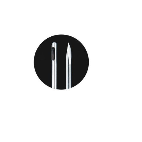
A microfracture awl allows the surgeon to create small holes in the subchondral bone without requiring a large drill pathway.
The instrument can be positioned directly against the prepared bone surface.
Controlled manual force is then used to create the perforation.
Potential practical considerations include:
Instrument tip geometry
Access angle
Working length
Surgeon control
Defect location
The objective is controlled penetration rather than extensive removal of subchondral bone.
Microfracture Awl vs Drilling: What Is the Difference?
Both techniques can be used to access the subchondral marrow, but they do so differently.
Microfracture Awl
An awl creates perforations by controlled impaction.
Drilling
A drill creates channels using a rotating cutting instrument.
The two approaches may differ in:
Hole geometry
Bone removal
Heat generation
Instrument control
Access angle
The chosen technique depends on the surgical strategy and surgeon preference.
Why Is the Cartilage Edge Prepared?
The damaged cartilage around the lesion may contain unstable tissue.
If this unstable tissue remains, the border of the defect may not provide a stable margin for the repair area.
The surgeon may therefore remove unstable cartilage and create stable vertical margins.
This helps define the lesion and provides a more controlled environment for the developing repair tissue.
Why Is the Calcified Cartilage Layer Important?
Between the articular cartilage and subchondral bone is a calcified cartilage layer.
Depending on the technique, this layer may be carefully removed or prepared before creating the microfracture holes.
The aim is to expose an appropriate subchondral surface while avoiding unnecessary damage to the underlying bone.
Precise defect preparation is therefore an important part of the procedure.
Why Is Rehabilitation Important After Microfracture?
The biological process continues long after surgery.
The repair tissue develops over time and is influenced by the mechanical environment.
For this reason, postoperative rehabilitation is a major component of microfracture treatment.
Depending on the defect, rehabilitation may include:
Temporary weight-bearing restrictions
Controlled range-of-motion exercises
Progressive strengthening
Gradual return to activity
The protocol depends strongly on defect location and associated procedures.
Why Might Weight Bearing Be Restricted?
A newly treated cartilage defect is mechanically vulnerable during the early healing period.
Excessive loading may disturb the developing repair tissue.
For defects on weight-bearing surfaces, the surgeon may therefore limit weight bearing for a period after surgery.
The duration and degree of restriction vary depending on:
Defect location
Defect size
Patient factors
Associated surgery
Surgeon protocol
Is Microfracture Used for Osteoarthritis?
Microfracture is generally associated with focal cartilage defects, not with widespread advanced osteoarthritis affecting the entire joint.
Diffuse degenerative cartilage loss represents a different biological and mechanical problem from a localized traumatic or focal chondral lesion.
Therefore, the presence of cartilage damage alone does not mean that microfracture is appropriate.
The complete joint condition should be considered.
Microfracture vs Osteochondral Grafting
Microfracture and osteochondral grafting use different principles.
Microfracture
Microfracture stimulates marrow-derived repair tissue.
Osteochondral Grafting
Osteochondral grafting transfers cartilage together with underlying bone into the defect.
Feature Microfracture Osteochondral Grafting
Main principle Marrow stimulation Tissue transplantation
Material added No cartilage graft Osteochondral graft
Repair tissue Marrow-derived Transferred cartilage/bone
Technique Subchondral perforation Graft harvesting/implantation
Defect selection Selected focal lesions Selected focal lesions
The appropriate method depends on lesion characteristics and overall treatment strategy.
Microfracture vs Autologous Chondrocyte Techniques
Cell-based cartilage procedures use a different biological strategy.
Microfracture relies on cells and biological factors released from the bone marrow.
Autologous chondrocyte-based techniques use cartilage cells that are collected and later implanted or incorporated into a repair construct.
These approaches differ in:
Biological source
Number of surgical stages
Technique complexity
Defect indications
Rehabilitation strategy
No single technique is appropriate for every cartilage lesion.
What Factors Can Affect Microfracture Outcomes?
Several factors can influence the biological and mechanical environment after treatment.
These may include:
Patient age
Defect size
Defect location
Body weight
Activity level
Limb alignment
Ligament stability
Meniscal integrity
Quality of surrounding cartilage
Previous cartilage surgery
Rehabilitation compliance
This demonstrates why cartilage treatment should be planned as part of the entire knee rather than focusing only on the visible defect.
Why Does Knee Alignment Matter?
If the mechanical axis of the lower limb places excessive load on the treated compartment, the cartilage defect may continue to experience high stress.
For example, malalignment can increase loading on one side of the knee.
In selected patients, cartilage treatment may therefore need to be considered together with correction of the mechanical environment.
Microfracture alone cannot correct abnormal limb alignment.
Why Do Meniscus and Ligament Stability Matter?
The menisci and ligaments influence load distribution and knee stability.
If the knee has:
Meniscal deficiency
ACL instability
PCL instability
Other ligament insufficiency
the cartilage surface may experience abnormal loading.
Therefore, associated pathology may need to be addressed together with the cartilage defect.
A stable mechanical environment is important for any cartilage repair strategy.
Can Microfracture Be Performed with Other Arthroscopic Procedures?
Yes.
Depending on the overall knee pathology, microfracture may be performed during the same surgical session as other arthroscopic procedures.
These may include:
Meniscal repair
Ligament reconstruction
Removal of loose bodies
Treatment of associated chondral lesions
Other arthroscopic interventions
The combined treatment strategy depends on the patient's complete pathology.
How Is Microfracture Different from Meniscal Repair?
These procedures treat completely different tissues.
Microfracture
Targets an articular cartilage defect and the underlying subchondral bone.
Meniscal Repair
Targets a tear in the meniscus.
Although both procedures can be performed arthroscopically, their biological goals and fixation principles are different.
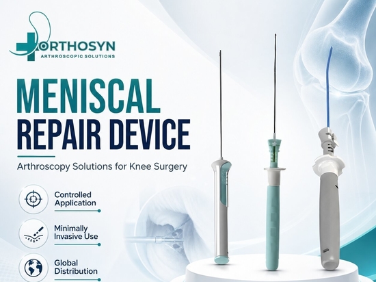
Orthosyn's arthroscopy portfolio includes solutions for both cartilage procedures and meniscal repair.
How Is Microfracture Different from ACL Reconstruction?
ACL reconstruction replaces or reconstructs a damaged ligament using a graft and fixation system.
Microfracture treats a focal cartilage defect through marrow stimulation.
The procedures may sometimes be performed in the same knee if both ligament instability and cartilage damage are present.
However, each addresses a different pathology.
What Are the Main Goals of Microfracture Surgery?
The principal goals are to:
Prepare a stable cartilage defect
Access the subchondral marrow
Create a biological healing response
Promote formation of repair tissue
Improve the local cartilage defect environment
The procedure does not guarantee restoration of native hyaline cartilage.
It is one cartilage repair strategy among several available options.
Orthosyn Microfracture Instrumentation
Orthosyn Medikal offers microfracture instrumentation as part of its arthroscopy and sports medicine portfolio.
The instruments are designed to support controlled arthroscopic access to the subchondral bone during selected cartilage repair procedures.
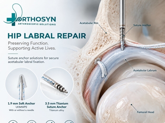
Orthosyn’s broader arthroscopy portfolio also includes:
Arthroscopic cannulas
Suture anchors
ACL/PCL reconstruction systems
Meniscal repair solutions
Passing instruments
Arthroscopic graspers
Other surgical instrumentation
These product groups support different stages of minimally invasive joint surgery.
Conclusion
Microfracture surgery is an arthroscopic marrow stimulation technique used for selected focal cartilage defects of the knee.
During the procedure, damaged and unstable cartilage is prepared and small perforations are created in the subchondral bone.
These perforations allow blood and marrow-derived biological components to enter the defect and form a clot that can develop into repair tissue.
The basic concept can be summarized as:
Cartilage Defect → Subchondral Perforation → Marrow Access → Biological Clot → Repair Tissue
Microfracture does not recreate normal hyaline cartilage directly and should not be considered appropriate for every type of cartilage damage.
Defect size, location, patient characteristics, alignment, meniscal function, ligament stability, rehabilitation, and the condition of the entire knee must all be considered when selecting a cartilage treatment strategy.
Orthosyn Medikal provides microfracture instrumentation designed to support controlled arthroscopic marrow stimulation procedures for selected cartilage defects.