#Product Trends
What Is Arthroscopic Broström Repair? | Orthosyn
Learn what chronic lateral ankle instability is, how the Broström repair restores the lateral ankle ligament complex, how arthroscopy is used during the procedure, and how suture anchors can support ligament reattachment to the fibula.
Arthroscopic Broström repair is a minimally invasive surgical technique used to treat selected patients with chronic lateral ankle instability, particularly when the lateral ankle ligaments have become insufficient after recurrent ankle sprains.
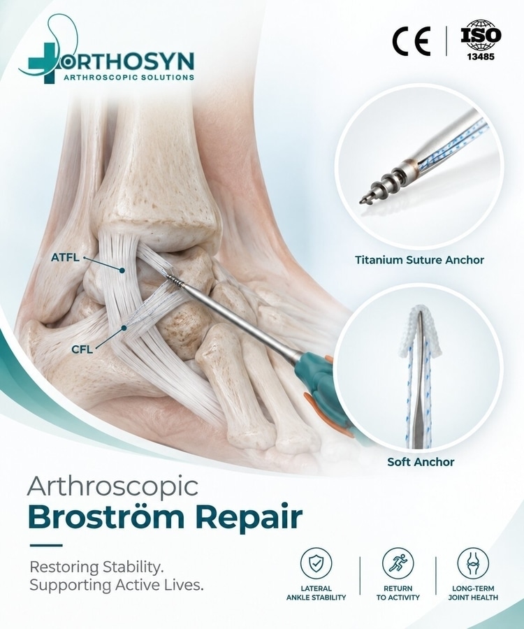
The traditional Broström procedure is based on anatomical repair of the injured lateral ankle ligaments, especially the anterior talofibular ligament (ATFL) and, when required, the calcaneofibular ligament (CFL). Modern arthroscopic techniques allow the surgeon to evaluate the ankle joint and perform selected components of ligament repair through small arthroscopic portals.
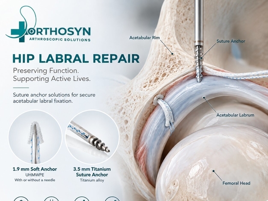
During an arthroscopic Broström repair, suture anchors can be inserted into the distal fibula to create fixation points. Sutures connected to the anchors are passed through the remaining ligament and capsular tissue and tensioned to restore attachment to the fibula.
The fundamental concept is:
Chronic Lateral Ankle Instability → Ligament Assessment → Fibular Preparation → Suture Anchor Placement → Ligament Reattachment → Ankle Stabilization
What Is Chronic Lateral Ankle Instability?
Ankle sprains are among the most common musculoskeletal injuries.
Many lateral ankle sprains involve the ligament structures on the outside of the ankle. Although many injuries can be managed non-operatively, repeated injuries or inadequate healing can result in persistent instability.
Patients with chronic lateral ankle instability may experience:
Recurrent ankle sprains
A sensation that the ankle is “giving way”
Difficulty during cutting or pivoting activities
Instability on uneven surfaces
Persistent lateral ankle discomfort
Reduced confidence during sports or physical activity
The condition can involve mechanical ligament insufficiency, functional instability, or a combination of both.
Which Ligaments Stabilize the Lateral Ankle?
The lateral ligament complex of the ankle primarily consists of three ligaments:
Anterior Talofibular Ligament — ATFL
Calcaneofibular Ligament — CFL
Posterior Talofibular Ligament — PTFL
Among these structures, the ATFL is particularly important in common inversion-type ankle injuries.
Anterior Talofibular Ligament
The ATFL extends between the distal fibula and talus.
It is frequently injured during lateral ankle sprains, particularly when the ankle is inverted in plantar flexion.
Calcaneofibular Ligament
The CFL connects the fibula to the calcaneus and contributes to lateral ankle and subtalar stability.
Depending on the injury pattern and degree of instability, both the ATFL and CFL may need to be considered during surgical stabilization.
Posterior Talofibular Ligament
The PTFL is a strong component of the lateral ligament complex and is generally less commonly injured in routine ankle sprains.
What Is a Broström Repair?
The Broström procedure is an anatomical ligament repair technique used to restore stability to the lateral ankle.
Rather than reconstructing the ligament complex with a tendon graft in every case, the procedure aims to repair the patient's remaining native ligament tissue when its quality is considered suitable.
The basic principle is:
Damaged Lateral Ligament → Anatomical Reattachment → Restoration of Lateral Ankle Stability
The procedure primarily addresses the ATFL and may also involve the CFL depending on the instability pattern and surgical technique.
What Is a Modified Broström Procedure?
The term modified Broström procedure is commonly used when the anatomical ligament repair is supplemented by additional reinforcement.
A frequently described modification is the Broström-Gould procedure, in which the repaired lateral ligament complex is reinforced using the inferior extensor retinaculum.
The objective remains anatomical stabilization while preserving and repairing the patient's native ligament structures.
Different variations exist, and the exact procedure depends on anatomy, tissue quality, instability pattern, surgeon preference, and associated pathology.
What Makes an Arthroscopic Broström Repair Different?
Traditional Broström repair is commonly performed using an open lateral ankle approach.
An arthroscopic Broström repair uses ankle arthroscopy to perform joint evaluation and selected repair steps through arthroscopic portals.
This can allow the surgeon to evaluate intra-articular pathology while addressing lateral ankle instability.
A general arthroscopic concept is:
Arthroscopic Evaluation → Identify Associated Pathology → Prepare Fibular Attachment Site → Place Suture Anchors → Capture Ligament/Capsule → Tension Repair
The exact technique can vary significantly between surgeons and fixation systems.
Why Is Arthroscopy Useful in Chronic Ankle Instability?
Chronic ankle instability may coexist with intra-articular abnormalities.
Arthroscopy allows direct visualization of the ankle joint and can help identify associated pathology such as:
Synovitis
Osteochondral lesions
Loose bodies
Cartilage abnormalities
Anterior impingement
Scar tissue
Other intra-articular abnormalities
This is one reason ankle arthroscopy may be incorporated into surgical management of chronic lateral ankle instability.
Not every associated finding requires treatment, and the surgical plan depends on the individual patient.
When Is Broström Repair Considered?
Broström repair may be considered in selected patients with symptomatic chronic lateral ankle instability when appropriate non-operative treatment has not provided sufficient stability.
Non-operative management may include:
Activity modification
Physiotherapy
Proprioceptive training
Strengthening
Bracing
Rehabilitation focused on neuromuscular control
Surgery is not automatically required after an ankle sprain.
The decision depends on symptoms, clinical instability, ligament quality, activity requirements, imaging findings, associated pathology, and response to conservative treatment.
How Is Chronic Lateral Ankle Instability Evaluated?
Evaluation generally combines patient history, physical examination, and imaging when appropriate.
Clinical assessment may include evaluation of:
Recurrent sprains
Giving-way episodes
Lateral ankle tenderness
Range of motion
Hindfoot alignment
Ligament laxity
Functional stability
Peroneal tendon function
Specific clinical tests can include the anterior drawer test and talar tilt test.
Imaging may include radiographs, MRI, ultrasound, or stress imaging depending on the clinical situation.
No single finding should necessarily be interpreted independently from the overall clinical assessment.
How Is Arthroscopic Broström Repair Performed?
The exact surgical technique varies, but a general procedure can include several stages.
1. Arthroscopic Evaluation
The arthroscope is introduced into the ankle joint through appropriate portals.
The surgeon evaluates the joint for associated abnormalities and assesses relevant ligament and capsular structures.
2. Preparation of the Fibular Attachment Site
The native attachment region of the lateral ligament complex on the distal fibula is identified.
The bone surface may be prepared to create an appropriate site for ligament reattachment.
3. Suture Anchor Placement
One or more suture anchors may be inserted into the distal fibula.
These anchors provide fixation points for the sutures used to capture and secure the ligamentous tissue.
4. Suture Passage
Sutures are passed through the ATFL and/or associated capsular tissue using an appropriate suture-passing technique.
5. Ligament Tensioning
The tissue is repositioned toward its anatomical attachment site.
Appropriate tension is important because the objective is to restore stability without unnecessarily restricting ankle motion.
6. Final Fixation
The sutures are secured according to the selected anchor system and surgical technique.
The surgeon evaluates the final repair and ankle stability.
How Are Suture Anchors Used in Broström Repair?
Suture anchors create fixation points between the lateral ligament tissue and the distal fibula.
The basic fixation concept is:
Distal Fibula → Suture Anchor → Suture → ATFL/Capsular Tissue
After the anchor is positioned in the fibula, the attached sutures can be passed through the ligamentous tissue.
The tissue is then brought back toward its anatomical attachment and secured.
This creates a bone-to-soft-tissue fixation construct intended to maintain the ligament in position during the healing process.
Why Is Anchor Position Important?
The distal fibula is a relatively small anatomical structure, and the anchor must be positioned with consideration for surrounding anatomy.
Important factors may include:
Anchor entry point
Drilling trajectory
Anchor diameter
Insertion depth
Bone quality
Distance between multiple anchors
Relationship to the articular surface
Available bone stock
Accurate instrumentation and controlled drilling are therefore important components of anchor-based ligament repair.
Why Does Suture Anchor Diameter Matter?
Every anchor requires a bone tunnel or insertion pathway.
Anchor diameter therefore influences the amount of bone required for implantation.
Smaller-diameter anchors may provide potential advantages in selected anatomical regions because they can require smaller bone tunnels.
This can be particularly relevant when:
The available bone stock is limited
Multiple anchors are required
Anchors must be positioned relatively close together
Bone preservation is important
Revision surgery may need to be considered
However, smaller does not automatically mean better.
Anchor selection should also consider fixation requirements, bone quality, implant design, suture configuration, surgical technique, and the dimensions of the anatomical region.
Can Soft All-Suture Anchors Be Used in Lateral Ankle Ligament Repair?
Soft all-suture anchors represent one category of anchor technology used in arthroscopic soft-tissue fixation.
Unlike conventional solid-body anchors, an all-suture anchor is primarily composed of high-strength suture material and is deployed within the bone after insertion through a relatively small tunnel.
Potential characteristics include:
Small insertion diameter
Reduced bone tunnel size
Low solid implant volume
Bone-preserving fixation concept
Ability to create multiple fixation points in limited anatomical areas
These characteristics can be relevant when planning soft-tissue fixation around smaller anatomical structures such as the distal fibula.
The suitability of an all-suture anchor for a specific procedure depends on the selected technique, bone quality, fixation requirements, implant specifications, and surgeon assessment.
What Is the Difference Between Soft and Solid Suture Anchors?
Suture anchors can be manufactured using different implant concepts and materials.
Feature Soft All-Suture Anchor Solid Suture Anchor
Implant concept Primarily suture-based Solid implant body
Bone tunnel Generally smaller Depends on anchor diameter
Implant volume Low solid implant volume Solid implant remains in bone
Fixation mechanism Deployment within bone Mechanical engagement of solid anchor
Bone preservation Important design consideration Depends on implant size and design
Application Soft-tissue fixation Soft-tissue fixation
Neither concept is universally preferable.
The appropriate anchor depends on the anatomy, bone quality, tissue characteristics, fixation strategy, and surgeon preference.
Can PEEK Suture Anchors Be Used for Soft-Tissue Fixation?
PEEK — polyether ether ketone — is a high-performance polymer used in orthopedic implants.
PEEK anchors can provide a solid-body fixation option and are commonly associated with characteristics such as:
Mechanical strength
Biocompatibility
Radiolucency
Resistance to degradation
Established use in orthopedic soft-tissue fixation
The choice between PEEK, titanium, all-suture, or another anchor technology depends on the procedure and specific fixation requirements.
What About Titanium Suture Anchors?
Titanium suture anchors are another established option for bone-to-soft-tissue fixation.
Potential characteristics include:
Solid metallic construction
Mechanical strength
Biocompatibility
Permanent implant structure
Long history of orthopedic use
As with other anchor technologies, appropriate anchor dimensions and positioning are important, particularly when working in anatomical regions with limited bone volume.
How Many Anchors Are Used in Broström Repair?
There is no universal number of anchors required for every Broström repair.
The number may depend on:
Extent of ligament injury
ATFL tissue quality
CFL involvement
Size of the distal fibula
Anchor dimensions
Bone quality
Repair configuration
Associated capsular laxity
Surgical technique
Some techniques may use a single fixation point, while others may use multiple anchors.
The number of anchors should therefore be determined by the requirements of the individual repair rather than by a fixed rule.
Is the ATFL Always the Only Ligament Repaired?
No.
The ATFL is frequently the primary ligament addressed in lateral ankle stabilization, but the injury pattern may involve additional structures.
The CFL may also contribute to instability, particularly when instability extends beyond isolated ATFL insufficiency.
The surgeon therefore evaluates the overall lateral ligament complex rather than assuming that every patient has an isolated ATFL injury.
What Is Ligament Tensioning and Why Is It Important?
After sutures have captured the ligament tissue, the tissue must be tensioned appropriately before final fixation.
Insufficient tension may leave residual instability.
Excessive tension may potentially alter normal ankle mechanics or restrict motion.
The objective is therefore not simply to make the ligament as tight as possible.
The goal is to restore appropriate anatomical tension and stability.
Ankle position during fixation can also be an important component of the selected surgical technique.
Broström Repair vs Ligament Reconstruction: What Is the Difference?
Broström repair and ligament reconstruction are related but different strategies.
Feature Broström Repair Ligament Reconstruction
Main concept Repair native ligament Reconstruct ligament using graft tissue
Native ATFL tissue Preserved and repaired when suitable May be insufficient for direct repair
Graft Usually not required for standard repair Commonly required
Suture anchors Commonly used May be used depending on technique
Primary objective Anatomical repair Replacement/reconstruction of deficient ligament
Selection Depends on tissue quality and instability Considered when direct repair may not be appropriate
Ligament reconstruction may be considered when native ligament tissue is inadequate for direct repair or when other patient-specific factors make reconstruction more appropriate.
What Factors Can Influence the Choice Between Repair and Reconstruction?
Several factors may influence surgical planning:
Quality of native ligament tissue
Severity and chronicity of instability
Previous ankle surgery
Generalized ligamentous laxity
Hindfoot alignment
Patient activity level
Body habitus
Associated injuries
Revision status
Surgeon assessment
Therefore, arthroscopic Broström repair should not be considered the appropriate procedure for every case of chronic lateral ankle instability.
What Is Internal Brace Augmentation?
Some lateral ankle stabilization procedures combine ligament repair with an additional high-strength suture-tape augmentation construct.
This is sometimes described as internal brace augmentation.
The concept is to provide supplementary mechanical support to the repaired ligament.
However, augmentation is not synonymous with a standard Broström repair and is not necessarily required in every patient.
Its use depends on the surgical strategy, patient characteristics, tissue quality, fixation system, and surgeon preference.
What Other Conditions May Be Evaluated During Ankle Arthroscopy?
One advantage of incorporating arthroscopy is the ability to inspect the ankle joint for associated pathology.
The surgeon may evaluate:
Osteochondral lesions of the talus
Synovitis
Anterior ankle impingement
Loose bodies
Cartilage lesions
Scar tissue
Joint surface abnormalities
The presence of these findings can influence the overall surgical plan.
What Instruments May Be Used During Arthroscopic Broström Repair?
Depending on the technique, instrumentation may include:
Arthroscope
Arthroscopic cannulas
Probes
Graspers
Shavers
Burrs
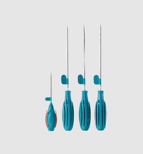
Suture anchors
Anchor drill guides
Drills
Suture passers
Tissue penetrators
Suture management instruments
Suture cutters
The exact instrumentation depends on the repair technique and fixation system.
What Should Be Considered When Selecting a Suture Anchor?
Suture anchor selection should be based on the complete fixation strategy.
Parameter Why It Matters
Anchor diameter Influences required bone tunnel size
Anchor material Determines implant characteristics
Distal fibular bone stock Influences available fixation options
Bone quality May affect fixation requirements
Anchor trajectory Important for controlled placement
Suture configuration Influences tissue capture
Number of anchors Determines repair configuration
Tissue quality Influences the overall repair strategy
Revision considerations May increase the importance of bone preservation
No single anchor design is ideal for every patient or every Broström technique.
Orthosyn Suture Anchor Solutions for Arthroscopic Soft-Tissue Repair
Orthosyn Medikal provides multiple suture anchor technologies for arthroscopic soft-tissue fixation, allowing different implant concepts to be considered according to anatomical dimensions, bone quality, fixation requirements, and surgical technique.
Relevant Orthosyn anchor technologies include:
Soft All-Suture Anchors
PEEK Suture Anchors
Titanium Suture Anchors
Knotless Anchor Systems
Multiple anchor diameters and suture configurations
Orthosyn Soft Suture Anchors
Orthosyn PEEK Suture Anchors
Orthosyn Titanium Suture Anchors
The selection of any anchor for lateral ankle ligament repair should be based on the dimensions of the implant, the selected surgical technique, bone quality, anatomical requirements, and the applicable product indications.
Conclusion
Arthroscopic Broström repair is a minimally invasive approach to anatomical lateral ankle ligament repair used in selected patients with chronic lateral ankle instability.
The procedure commonly focuses on repairing the anterior talofibular ligament (ATFL) and may also address the calcaneofibular ligament (CFL) depending on the instability pattern.
A simplified repair concept is:
Chronic Ankle Instability → Arthroscopic Evaluation → Fibular Preparation → Suture Anchor Placement → Suture Passage → Controlled Ligament Tensioning → Anatomical Fixation
Suture anchors provide fixation points in the distal fibula that allow the remaining ligament and capsular tissue to be reattached. Anchor diameter, implant design, bone quality, drilling trajectory, suture configuration, ligament quality, and repair tension are all relevant to the overall fixation strategy.
Arthroscopy also allows the surgeon to evaluate associated intra-articular abnormalities that may coexist with chronic lateral ankle instability.
The appropriate surgical approach depends on the patient's anatomy, ligament quality, instability pattern, associated pathology, previous treatment, and surgeon assessment.
Orthosyn Medikal offers soft all-suture, PEEK, titanium, and knotless suture anchor technologies designed to support different arthroscopic soft-tissue fixation strategies where the selected implant dimensions, fixation characteristics, surgical technique, and applicable indications are appropriate.