#Product Trends
How Are Suture Anchors Used in TFCC Repair? | Orthosyn
Learn how suture anchors can be used during arthroscopic triangular fibrocartilage complex repair to reattach selected peripheral TFCC tears to the ulnar attachment, restore soft-tissue fixation, and support distal radioulnar joint stability.
The triangular fibrocartilage complex (TFCC) is an important stabilizing structure on the ulnar side of the wrist. It contributes to the stability of the distal radioulnar joint (DRUJ) and helps transmit and distribute forces between the hand, wrist, radius, and ulna.
TFCC injuries can occur following trauma, repetitive loading, rotational forces, or degenerative changes. Depending on the location and characteristics of the tear, treatment may range from conservative management to arthroscopic debridement or surgical repair.
For selected repairable peripheral TFCC tears, suture anchors can be used to secure the injured tissue back to its anatomical attachment.
The basic concept can be represented as:
TFCC Tear → Arthroscopic Assessment → Preparation of Ulnar Attachment → Suture Anchor Placement → Suture Passage Through TFCC → Tissue Reattachment → Stabilization
The purpose of the anchor is not to replace the TFCC. Instead, it provides a fixation point in bone that allows sutures to secure the repairable soft tissue while biological healing develops.
What Is the TFCC?
The triangular fibrocartilage complex is a group of interconnected soft-tissue structures located on the ulnar side of the wrist.
It includes structures commonly described as:
- Triangular fibrocartilage disc
- Dorsal radioulnar ligament
- Volar radioulnar ligament
- Ulnolunate ligament
- Ulnotriquetral ligament
- Meniscus homologue
- Ulnar-sided capsular structures
The TFCC functions as both a load-transmitting structure and a stabilizing complex.
One of its particularly important roles is stabilization of the distal radioulnar joint during forearm rotation.
What Is the Distal Radioulnar Joint?
The distal radioulnar joint is formed between the distal radius and ulna near the wrist.
It allows the radius to rotate around the ulna during:
- Pronation
- Supination
These movements are essential for activities such as turning a key, using a screwdriver, opening a door handle, or rotating the palm upward and downward.
The TFCC and particularly its radioulnar ligament components contribute to DRUJ stability throughout this rotational movement.
A significant TFCC injury can therefore be associated with:
TFCC Damage → Reduced Soft-Tissue Restraint → DRUJ Instability → Pain and Functional Limitation
However, not every TFCC tear produces clinically significant DRUJ instability.
What Causes a TFCC Tear?
TFCC injuries may be traumatic or degenerative.
Traumatic TFCC Tears
Potential mechanisms include:
- Falling onto an outstretched hand
- Forceful wrist rotation
- Sports injuries
- Axial loading of the wrist
- Sudden twisting injuries
- High-energy trauma
Degenerative TFCC Tears
Degenerative changes may develop gradually and can be associated with repetitive loading, aging, ulnar-sided wrist mechanics, and other anatomical or mechanical factors.
The mechanism and location of the lesion influence whether repair is appropriate.
What Are the Symptoms of a TFCC Injury?
Possible symptoms include:
- Ulnar-sided wrist pain
- Pain during forearm rotation
- Pain during gripping
- Clicking or catching
- Reduced grip strength
- Tenderness on the ulnar side of the wrist
- Sensation of instability
- Pain during weight-bearing through the wrist
These findings are not specific to TFCC tears.
Other causes of ulnar-sided wrist pain may produce similar symptoms, so clinical examination and appropriate imaging are important.
How Are TFCC Tears Evaluated?
Assessment may include:
- Clinical history
- Physical examination
- DRUJ stability assessment
- Wrist radiographs
- MRI
- MR arthrography in selected situations
- Diagnostic wrist arthroscopy
Arthroscopy can provide direct visualization of the TFCC and surrounding intra-articular structures.
It may also allow the surgeon to assess:
- Tear location
- Tissue quality
- Stability
- Repairability
- Associated cartilage lesions
- Other intra-articular abnormalities
Does Every TFCC Tear Require Surgery?
No.
Many TFCC injuries can initially be managed non-operatively depending on the lesion and clinical situation.
Conservative management may include:
- Activity modification
- Immobilization or bracing
- Rehabilitation
- Medical management when clinically appropriate
Surgical treatment may be considered when symptoms persist, instability is clinically important, or the tear has characteristics that make surgical treatment appropriate.
Importantly, not every TFCC tear should be repaired with a suture anchor.
Treatment depends heavily on tear location, tissue quality, vascularity, DRUJ stability, associated pathology, patient characteristics, and surgical assessment.
Why Does the Location of a TFCC Tear Matter?
Different regions of the TFCC have different biological and mechanical characteristics.
Peripheral regions generally have better vascularity than the central portion.
This distinction is important because healing potential is one of the factors considered when determining whether a tear is suitable for repair.
Conceptually:
Peripheral Repairable Tear → Potential for Tissue Reattachment
while
Central Poorly Vascularized Tear → Repair May Not Be the Preferred Strategy
This is a simplified distinction. Actual treatment depends on the complete injury pattern.
What Is Arthroscopic TFCC Repair?
Arthroscopic TFCC repair is a minimally invasive surgical approach in which a small arthroscope and specialized instruments are introduced into the wrist.
The surgeon can inspect the TFCC directly and determine the characteristics of the lesion.
When repair is appropriate, the procedure may involve:
Arthroscopic Evaluation → Tear Identification → Tissue Preparation → Fixation → Stability Assessment
Different repair techniques exist.
These can include:
- Inside-out repair
- Outside-in repair
- All-inside repair
- Suture anchor repair
- Foveal repair techniques
- Capsular repair techniques
The appropriate method depends on the location and pattern of the TFCC injury.
Why Are Suture Anchors Used in TFCC Repair?
A suture anchor creates a fixation point between bone and suture.
The anchor is inserted into bone, while the attached suture is passed through the repairable TFCC tissue.
The sutures are then used to bring the tissue toward its intended attachment.
The basic principle is:
Bone → Suture Anchor → Suture → TFCC Tissue
This converts the bone into a stable fixation point for soft-tissue reattachment.
Where Can a Suture Anchor Be Placed During TFCC Repair?
Anchor position depends on the tear pattern and repair technique.
For selected ulnar-sided or foveal TFCC injuries, fixation may involve the distal ulna.
A simplified concept is:
Ulnar Attachment → Anchor Placement → TFCC Capture → Tissue Reattachment
Precise positioning is important because the ulnar side of the wrist contains multiple anatomical structures within a relatively small area.
Anchor trajectory, implant diameter, bone stock, tissue location, and surrounding anatomy therefore require careful consideration.
What Is a Foveal TFCC Tear?
The TFCC has important deep attachment fibers at the ulnar foveal region.
These deep fibers contribute significantly to DRUJ stability.
When the foveal attachment is disrupted, the TFCC may lose part of its stabilizing connection to the ulna.
In selected cases, repair may therefore focus on restoring this attachment.
The concept is:
Foveal Detachment → Preparation of Ulnar Fixation Site → Suture Fixation → Reattachment of TFCC
Suture anchors represent one possible fixation strategy for this type of repair.
Why Is Foveal Repair Important for DRUJ Stability?
The deep radioulnar ligament components associated with the foveal attachment are important stabilizers of the DRUJ.
If these fibers are detached, instability may occur depending on the extent and pattern of injury.
A foveal repair attempts to restore the soft-tissue attachment responsible for this stabilizing function.
The objective is therefore not merely to close a visible tear.
It is to restore the relevant anatomical attachment when clinically indicated.
How Is a Suture Anchor Inserted?
The exact technique varies according to the implant system and surgical approach.
In general, anchor-based fixation can involve:
Bone Preparation → Anchor Insertion → Suture Passage → Tissue Reduction → Suture Fixation
The surgeon first identifies the intended fixation site.
Depending on the anchor design, a pilot hole or bone preparation may be required.
The anchor is then inserted at a controlled trajectory.
The attached sutures are passed through the selected TFCC tissue and secured according to the repair technique.
Why Does Anchor Diameter Matter in TFCC Repair?
The distal ulna and the anatomical structures around the wrist provide a relatively limited working area compared with larger joints such as the shoulder.
Anchor diameter can therefore be an important consideration.
Smaller anchor systems may allow a smaller bone tunnel or socket.
Conceptually:
Smaller Anchor Diameter → Smaller Bone Preparation → Potential for Greater Bone Preservation
However, smaller is not automatically better.
Anchor selection must also consider:
- Required fixation
- Bone quality
- Tissue characteristics
- Implant design
- Surgical technique
- Available bone stock
- Anchor trajectory
The objective is to balance fixation requirements with preservation of the available bone.
Why Can Soft Suture Anchors Be Relevant?
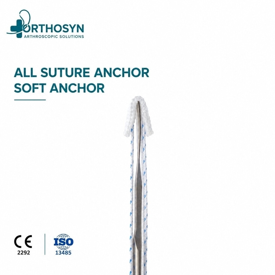
Soft or all-suture anchors use a predominantly suture-based anchoring component rather than a conventional rigid anchor body.
Because these systems can be available in small diameters, they may be useful when bone preservation and a small fixation footprint are important.
Potential technical characteristics include:
- Small bone preparation
- Reduced rigid implant volume
- Soft-tissue fixation capability
- Availability in small diameters
For a small anatomical region such as the wrist, these characteristics may be relevant when compatible with the required fixation and surgical technique.
However, the selection of an all-suture anchor depends on the specific procedure, bone quality, fixation requirements, implant dimensions, applicable indications, and surgeon preference.
Can PEEK Anchors Be Used for Soft-Tissue Fixation?
PEEK is a high-performance polymer used in various orthopedic implants.
PEEK suture anchors provide a rigid implant body and can be used for bone-to-soft-tissue fixation in procedures where their dimensions and design are appropriate.
Characteristics associated with PEEK include:
- Biocompatibility
- Radiolucency
- Mechanical stability
- Resistance to degradation
- Permanent polymer construction
Whether a PEEK anchor is suitable for a specific TFCC technique depends on implant dimensions, available bone, surgical approach, and the product's applicable indications.
Can Titanium Suture Anchors Be Used?
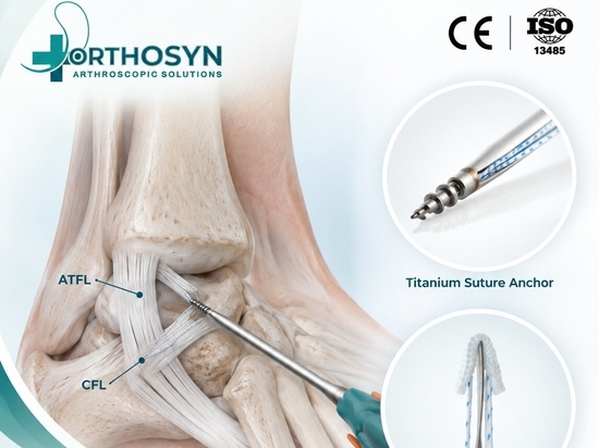
Titanium suture anchors are another established category of bone-to-soft-tissue fixation devices.
Potential characteristics include:
- Metallic construction
- Mechanical strength
- Biocompatibility
- Permanent fixation
- Established use in orthopedic soft-tissue fixation
As with PEEK and soft anchors, titanium anchor selection depends on the anatomy, anchor dimensions, bone stock, surgical technique, and applicable indications.
There is no single anchor material that is universally preferable for every TFCC repair.
Soft vs PEEK vs Titanium Anchors
Anchor Type General Characteristic Potential Consideration
Soft / All-Suture Predominantly suture-based anchor body Small bone preparation and bone preservation
PEEK Rigid polymer anchor Radiolucent permanent implant
Titanium Metallic anchor Rigid permanent metallic fixation
Material alone does not determine whether an anchor is appropriate.
The diameter, geometry, fixation mechanism, suture configuration, bone quality, anatomical location, and surgical technique must also be considered.
How Are Sutures Passed Through the TFCC?
After anchor placement, the attached suture must capture sufficient repairable TFCC tissue.
Depending on the technique, specialized instruments may be used to pass the suture through the tissue.
These can include:
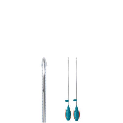
- Suture passers
- Small arthroscopic graspers
- Needles
- Cannulas
- Suture retrieval instruments
The objective is to create a secure tissue purchase while respecting the surrounding anatomy.
Why Is Suture Placement Important?
Anchor fixation is only one component of the repair.
The suture must capture the appropriate tissue.
Poor tissue capture may compromise the repair even when the anchor itself is securely fixed in bone.
The complete fixation construct can therefore be considered as:
Bone Fixation + Anchor Stability + Suture Integrity + Tissue Capture + Appropriate Tension
All components contribute to the overall repair.
How Much Tension Should Be Applied?
The objective of repair is generally to restore the tissue toward its anatomical attachment without inappropriate overtensioning.
Tension depends on:
- Tear pattern
- Tissue quality
- Repair location
- Suture configuration
- Anchor position
- DRUJ stability
- Surgical technique
Excessive tension can alter tissue mechanics, while insufficient tension may fail to restore the intended attachment.
Therefore, fixation should be evaluated as part of the complete repair construct.
Knotted vs Knotless Fixation in TFCC Repair
Suture anchor systems may use knotted or knotless fixation concepts.
Knotted Fixation
The surgeon passes the suture through the tissue and creates arthroscopic or surgical knots to secure the repair.
Knotless Fixation
A knotless system secures the suture without requiring a conventional final knot.
Potential considerations can include:
- Surgical access
- Suture management
- Implant dimensions
- Fixation mechanism
- Tissue tensioning
- Surgeon preference
Neither concept is universally superior for every TFCC repair.
The technique must be selected according to the specific anatomy and repair strategy.
How Many Suture Anchors Are Required?
There is no universal number.
The number of anchors depends on factors such as:
- Tear size
- Tear location
- Repair configuration
- Tissue quality
- Required fixation
- Anchor design
- Available bone
- Surgical technique
Some repairs may require a single fixation point, while other configurations may require additional fixation.
The objective is appropriate anatomical repair rather than maximizing the number of implants.
Why Is Bone Preservation Important in Wrist Surgery?
The wrist contains relatively small bones and compact anatomical regions.
Preserving bone stock may therefore be especially relevant.
The amount of bone preparation depends on:
- Anchor diameter
- Drill diameter
- Implant design
- Number of anchors
- Spacing between fixation points
- Bone quality
This is one reason small-diameter anchor technologies can be useful in selected wrist procedures.
What Structures Must Be Considered During TFCC Repair?
The ulnar side of the wrist contains important structures that must be considered during surgical planning and repair.
These include:
- Distal ulna
- DRUJ
- Extensor carpi ulnaris region
- Ulnar-sided neurovascular structures
- Articular cartilage
- Surrounding capsular and ligamentous structures
Precise portal placement, drilling trajectory, suture passage, and implant positioning are therefore important.
Is TFCC Repair the Same as TFCC Debridement?
No.
These procedures address different types of pathology.
TFCC Debridement
Debridement removes unstable or damaged tissue that is not being anatomically reattached.
It may be considered for selected lesions, including certain central tears.
TFCC Repair
Repair attempts to preserve and reattach repairable TFCC tissue.
Conceptually:
Debridement → Removal of Unstable Damaged Tissue
Repair → Preservation + Reattachment of Repairable Tissue
The choice depends on tear location, tissue quality, vascularity, stability, associated pathology, and clinical assessment.
What Instruments May Be Used During Arthroscopic TFCC Repair?
A TFCC repair procedure may involve:
- Wrist arthroscope
- Small-diameter arthroscopic instruments
- Probe
- Graspers
- Suture passers
- Needles
- Cannulas
- Drills or bone preparation instruments
- Suture anchors
- Suture management instruments
Instrument dimensions are particularly important because wrist arthroscopy is performed within a small joint space.
What Factors Influence Suture Anchor Selection?
Parameter Why It Matters
Anchor diameter Influences bone preparation and fixation footprint
Anchor material Determines implant characteristics
Bone quality Influences anchor-bone interaction
Tear location Determines fixation strategy
Tissue quality Influences suture purchase
Suture configuration Affects tissue capture and repair construct
Anchor trajectory Influences positioning and surrounding structures
Available bone stock Important in small anatomical regions
Knotted vs knotless design Influences fixation technique
Surgical approach Determines access and implant requirements
Anchor selection should therefore be based on the complete repair strategy rather than on one implant characteristic alone.
Orthosyn Suture Anchor Solutions for Arthroscopic Soft-Tissue Repair
Orthosyn Medikal provides several suture anchor technologies for arthroscopic bone-to-soft-tissue fixation, including:
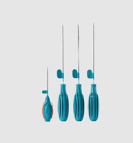
Orthosyn Soft / All-Suture Anchors
Small-diameter soft anchor systems can be relevant when minimizing bone preparation is an important technical consideration.
Orthosyn Soft Anchors
Orthosyn PEEK Suture Anchors
PEEK anchor systems provide rigid polymer-based bone-to-soft-tissue fixation options.
Orthosyn PEEK Suture Anchors
Orthosyn Titanium Suture Anchors
Titanium anchor systems provide metallic fixation options for arthroscopic soft-tissue repair.
Orthosyn Titanium Suture Anchors
Orthosyn Knotless Anchor Systems
Knotless anchor technologies provide an alternative fixation concept when knotless tissue fixation is compatible with the selected procedure and technique.
Orthosyn PEEK + PEEK Knotless Anchor System
Orthosyn PEEK + Titanium Knotless Anchor
The suitability of any specific Orthosyn anchor for TFCC repair depends on the implant dimensions, fixation requirements, surgical technique, applicable product indications, and clinical assessment.
Conclusion
Suture anchors can be used in selected TFCC repairs to create a bone-based fixation point for reattachment of repairable soft tissue.
The basic concept is:
TFCC Tear → Arthroscopic Evaluation → Identification of Repairable Tissue → Ulnar Fixation Site Preparation → Suture Anchor Placement → Suture Passage → TFCC Reattachment → Stability Assessment
For selected peripheral or foveal injuries, anchor-based fixation may allow the surgeon to secure TFCC tissue toward its ulnar attachment. This can be particularly relevant when restoration of the stabilizing soft-tissue attachment is part of the surgical objective.
Successful repair depends on much more than the anchor itself. Important factors include tear location, tissue quality, DRUJ stability, vascularity, bone quality, anchor diameter, fixation mechanism, suture placement, tension, surgical trajectory, and rehabilitation.
Soft all-suture, PEEK, titanium, and knotless anchor technologies provide different fixation characteristics. No single anchor type is universally appropriate for every TFCC injury.
Orthosyn Medikal provides soft all-suture, PEEK, titanium, and knotless suture anchor technologies for arthroscopic bone-to-soft-tissue fixation, offering different implant concepts and dimensions for procedures where the selected anchor, surgical technique, anatomical requirements, and applicable indications are appropriate.