#Product Trends
How Is the Subscapularis Tendon Repaired Arthroscopically? | Orthosyn
Learn how arthroscopic subscapularis tendon repair is performed, how suture anchors are used to reattach the tendon to the lesser tuberosity, and why tendon mobilization, anatomical footprint preparation, suture configuration, and fixation stability
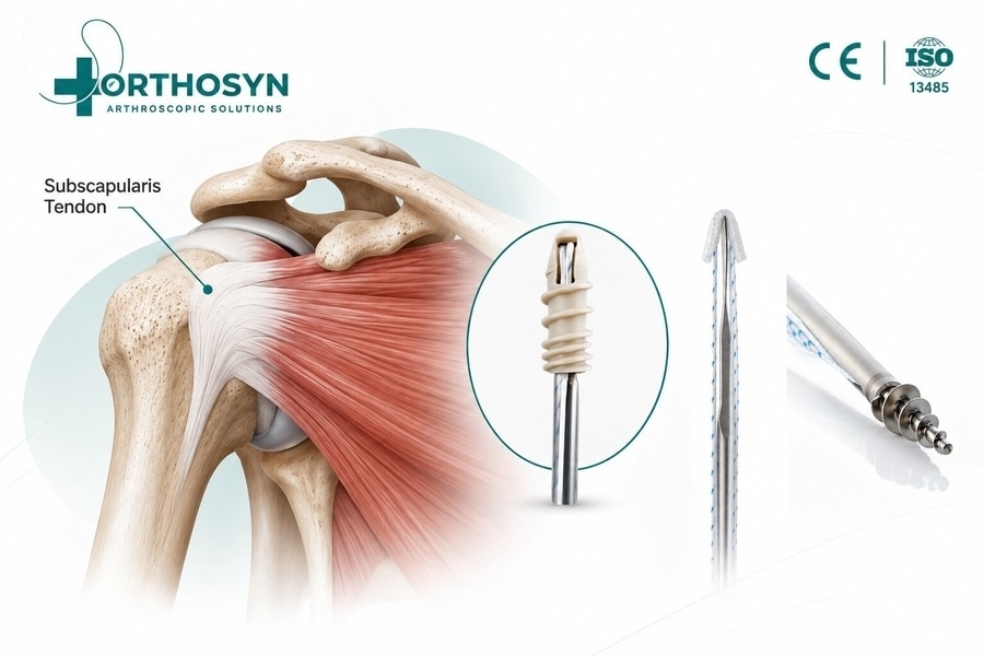
The subscapularis tendon is an essential component of the rotator cuff and plays an important role in shoulder stability, internal rotation, and coordinated glenohumeral movement.
When the subscapularis tendon is torn, patients may experience shoulder pain, weakness, difficulty performing internal rotation, and reduced functional stability. Depending on the severity of the injury, tendon quality, patient characteristics, and associated shoulder conditions, arthroscopic surgical repair may be considered.
Arthroscopic subscapularis repair is a minimally invasive surgical procedure designed to reattach the damaged tendon to its anatomical insertion on the lesser tuberosity of the humerus.
During the procedure, surgeons may use suture anchors to establish fixation points within the bone. High-strength sutures are passed through the tendon and secured to maintain contact between the tendon and its prepared attachment site.
The fundamental concept is:
Subscapularis Tendon Tear → Arthroscopic Assessment → Tendon Mobilization → Footprint Preparation → Suture Anchor Placement → Tendon Reattachment → Biological Healing
Modern arthroscopic techniques may involve titanium, PEEK, soft all-suture, or knotless anchor systems, depending on the tear configuration, bone quality, surgical technique, and applicable implant indications.
Within the Orthosyn Medikal arthroscopy portfolio, different suture anchor technologies provide fixation options for selected arthroscopic soft-tissue repair procedures.
What Is the Subscapularis Tendon?
The subscapularis is one of the four muscles forming the rotator cuff.
The rotator cuff consists of:
- Supraspinatus
- Infraspinatus
- Teres minor
- Subscapularis
The subscapularis muscle originates from the anterior surface of the scapula and inserts primarily onto the lesser tuberosity of the humerus.
Its main functions include:
- Internal rotation of the shoulder
- Dynamic stabilization of the humeral head
- Contribution to anterior shoulder stability
- Maintenance of balanced shoulder movement
- Coordination with other rotator cuff muscles
The subscapularis is particularly important because it forms a major part of the anterior rotator cuff.
What Is a Subscapularis Tendon Tear?
A subscapularis tendon tear occurs when part or all of the tendon becomes damaged or detached from its humeral attachment.
The injury may involve:
- Partial-thickness tearing
- Full-thickness tearing
- Superior tendon tears
- Larger tears extending inferiorly
- Complete tendon detachment
- Retracted tendon tears
Subscapularis tears can occur independently or together with other rotator cuff injuries.
They are frequently associated with abnormalities involving the long head of the biceps tendon and its stabilizing pulley structures.
What Causes Subscapularis Tendon Tears?
Subscapularis injuries can develop through traumatic or degenerative mechanisms.
Potential causes include:
- Falls onto an outstretched arm
- Forced external rotation injuries
- Shoulder dislocation
- Sports-related trauma
- Repetitive mechanical loading
- Age-related tendon degeneration
- Chronic rotator cuff disease
Acute traumatic tears may involve sudden tendon detachment, while degenerative tears can develop progressively.
The mechanism of injury can influence tendon quality, retraction, associated pathology, and the surgical strategy.
What Are the Symptoms of a Subscapularis Tear?
Patients with subscapularis tendon injuries may experience:
- Anterior shoulder pain
- Weakness during internal rotation
- Difficulty reaching behind the back
- Reduced shoulder strength
- Pain during lifting or pushing
- Functional limitations
- Discomfort during certain rotational movements
Some patients may also have associated biceps tendon symptoms.
However, symptoms alone cannot reliably determine the precise tear pattern.
Clinical examination and appropriate imaging are important.
How Is a Subscapularis Tear Diagnosed?
Diagnosis typically involves a combination of clinical examination and imaging.
Clinical Examination
Commonly used clinical tests include:
- Lift-off test
- Belly-press test
- Bear-hug test
- Internal rotation strength assessment
These tests help evaluate subscapularis function, although their diagnostic performance varies with tear size and patient characteristics.
Imaging
Magnetic resonance imaging (MRI) is commonly used to evaluate:
- Tendon integrity
- Tear size
- Tendon retraction
- Muscle atrophy
- Fatty infiltration
- Associated rotator cuff tears
- Biceps pulley abnormalities
Ultrasound may also be useful when performed by an experienced operator.
Arthroscopy provides direct visualization of the tendon and associated intra-articular structures.
Does Every Subscapularis Tear Require Surgery?
No.
Not every subscapularis tendon tear requires surgical treatment.
Management depends on:
- Tear size
- Partial or full-thickness involvement
- Traumatic or degenerative origin
- Patient age
- Functional requirements
- Symptoms
- Tendon quality
- Degree of retraction
- Muscle condition
- Associated shoulder pathology
- Response to conservative treatment
Selected partial tears and lower-demand clinical situations may be managed without surgery.
However, acute traumatic full-thickness tears, progressive weakness, or symptomatic repairable tears may warrant surgical evaluation.
The treatment decision should be individualized.
What Is Arthroscopic Subscapularis Tendon Repair?
Arthroscopic subscapularis tendon repair is a minimally invasive procedure performed through small portals using an arthroscope and specialized instruments.
The objective is to restore the tendon’s attachment to the lesser tuberosity.
The surgeon evaluates the tear, mobilizes the tendon when necessary, prepares the bone attachment site, and secures the tendon using an appropriate fixation technique.
Suture anchors are commonly used to create fixation points in the humerus.
The basic fixation concept is:
Lesser Tuberosity → Suture Anchor → High-Strength Suture → Subscapularis Tendon
The anchor provides mechanical fixation while biological tendon-to-bone healing develops.
Why Is the Lesser Tuberosity Important?
The lesser tuberosity is the primary bony insertion region of the subscapularis tendon.
During repair, the surgeon aims to restore appropriate contact between the torn tendon and its anatomical attachment area.
This attachment region is commonly referred to as the tendon footprint.
The footprint is important because it influences:
- Anatomical tendon positioning
- Tendon-to-bone contact
- Load distribution
- Repair tension
- Biological healing conditions
The objective is not simply to attach the tendon somewhere on the humerus.
Instead, the repair should restore the tendon as closely as practical to its appropriate anatomical insertion.
How Is Arthroscopic Subscapularis Repair Performed?
The general procedure can be summarized as:
1. Arthroscopic shoulder assessment
Evaluate the tendon tear and associated shoulder pathology
2. Tendon identification and mobilization
Assess tissue mobility and release adhesions when necessary
3. Lesser tuberosity footprint preparation
Prepare the tendon attachment site while preserving viable bone
4. Suture anchor placement
Establish fixation points according to the repair configuration
5. Suture passage and tendon fixation
Restore tendon contact with the prepared footprint
6. Final repair assessment
Evaluate tendon position, tension, fixation, and stability
The exact sequence and technique vary depending on the tear and surgeon's approach.
Step 1: Arthroscopic Assessment of the Shoulder
The procedure begins with arthroscopic examination of the shoulder joint.
The surgeon evaluates the subscapularis tendon and surrounding structures.
Important findings may include:
- Tear location
- Tear size
- Tendon retraction
- Tissue quality
- Remaining tendon attachment
- Biceps tendon position
- Rotator interval abnormalities
- Associated supraspinatus injuries
- Cartilage or labral pathology
The superior portion of the subscapularis tendon deserves particular attention because smaller tears may be difficult to identify during routine assessment.
A thorough evaluation helps determine whether repair is appropriate and what fixation strategy may be required.
Step 2: Identification and Mobilization of the Torn Tendon
After identifying the tear, the surgeon evaluates tendon mobility.
Some tears can be reduced to the lesser tuberosity without extensive mobilization.
Others, particularly chronic or retracted tears, may require controlled release of adhesions and surrounding tissue restrictions.
The goal is to achieve sufficient tendon mobility for anatomical reduction without excessive tension.
Conceptually:
Tendon Retraction → Controlled Mobilization → Improved Footprint Access → Reduced Repair Tension
However, excessive tissue release can damage important structures.
The extent of mobilization depends on anatomy, tear configuration, tissue condition, and surgical judgment.
Why Is Tendon Mobilization Important?
A tendon that cannot reach its anatomical footprint without excessive tension may be difficult to repair reliably.
Excessive tension can increase mechanical stress on:
- Sutures
- Suture-tendon interfaces
- Anchor fixation
- Tendon tissue
- Tendon-to-bone healing interface
Therefore, successful repair requires more than strong implants.
It also requires appropriate tissue positioning.
Adequate Tendon Mobility + Anatomical Reduction + Controlled Tension = Important Conditions for Repair
Step 3: Preparation of the Lesser Tuberosity Footprint
The surgeon prepares the lesser tuberosity attachment site to support tendon-to-bone healing.
This may involve removing residual soft tissue and preparing the bone surface according to the surgical technique.
The objective is to establish an appropriate healing interface while preserving sufficient bone for anchor fixation.
Footprint preparation should balance:
- Biological healing conditions
- Bone preservation
- Anchor stability
- Anatomical positioning
- Avoidance of unnecessary bone removal
Excessive bone removal is not required simply to create a fixation surface.
Step 4: Placement of Suture Anchors
Suture anchors are inserted into the humerus at positions selected to support the repair configuration.
Their role is to establish stable fixation points for the sutures that secure the subscapularis tendon.
The fixation relationship can be summarized as:
Humeral Bone → Suture Anchor → Suture → Tendon → Anatomical Reattachment
Anchor selection depends on several factors:
- Tear size
- Tendon quality
- Bone quality
- Anchor diameter
- Anchor material
- Available fixation area
- Suture configuration
- Surgeon preference
- Applicable product indications
No single anchor type is universally appropriate for every subscapularis repair.
Why Does Anchor Position Matter?
Correct anchor placement helps establish the desired tendon-to-bone contact.
Improper placement may contribute to:
- Non-anatomical tendon positioning
- Uneven tension
- Inadequate footprint coverage
- Unfavorable suture direction
- Reduced fixation efficiency
- Potential interference with surrounding structures
Anchor positioning must be planned according to the anatomy of the lesser tuberosity and the intended repair.
The goal is not simply to place an anchor securely in bone, but to position it so that the suture construct supports appropriate tendon reduction.
Why Does Anchor Diameter Matter?
Anchor diameter influences the amount of bone removed during preparation and implantation.
A smaller anchor may require a smaller bone hole, potentially preserving more bone.
However, smaller diameter does not automatically mean superior fixation.
Anchor selection must consider:
- Bone quality
- Fixation strength requirements
- Implant geometry
- Suture configuration
- Available bone stock
- Surgical technique
Conceptually:
Smaller Required Bone Hole → Less Bone Removed
But:
Smaller Anchor ≠ Automatically Stronger or Better Repair
The objective is to balance fixation requirements with bone preservation.
Step 5: Passing Sutures Through the Subscapularis Tendon
After anchor placement, sutures are passed through the tendon using appropriate arthroscopic instruments.
Suture passage is a critical stage because the configuration determines how fixation forces are transmitted through the tendon.
Possible techniques may include:
- Simple sutures
- Mattress sutures
- Modified mattress configurations
- Rip-stop constructs
- Other technique-specific patterns
The selected configuration depends on tendon quality, tear size, repair strategy, and surgeon preference.
Why Is Suture Configuration Important?
The suture configuration influences how mechanical loads are distributed across the repaired tendon.
Poor tissue quality may increase the risk of sutures cutting through the tendon.
For this reason, surgeons may select configurations intended to improve tissue capture or distribute loads across a larger area.
The general principle is:
Suture Configuration → Tissue Capture → Load Distribution → Tendon Fixation
However, no single suture configuration is universally superior for all tear patterns.
Step 6: Reattaching the Tendon to the Lesser Tuberosity
Once sutures have been passed through the tendon, they are secured to bring the tendon into contact with the prepared footprint.
Depending on the implant and technique, fixation may involve:
- Arthroscopic knot tying
- Knotless fixation
- Single-row repair
- Double-row repair
- Hybrid fixation strategies
The objective is to restore tendon position while avoiding excessive tension.
Anatomical Tendon Reduction → Controlled Suture Tension → Footprint Contact → Mechanical Fixation
Mechanical fixation provides initial stability.
Long-term biological integration depends on tendon-to-bone healing.
What Is Single-Row Subscapularis Repair?
Single-row repair generally uses one row of anchors to secure the tendon to the lesser tuberosity.
It may be considered for selected tear patterns where appropriate tendon reduction and fixation can be achieved.
Potential characteristics include:
- Relatively straightforward fixation configuration
- Fewer fixation points in some techniques
- Direct tendon-to-bone reattachment
- Compatibility with knotted or knotless constructs
The suitability of single-row repair depends on the tear and surgical requirements.
What Is Double-Row Subscapularis Repair?
Double-row repair uses a fixation strategy involving medial and lateral fixation points to support tendon-to-bone contact.
Depending on the technique, it may provide a broader footprint compression pattern.
However, double-row repair is not automatically required for every subscapularis tear.
Important considerations include:
- Tear dimensions
- Tendon mobility
- Tissue quality
- Footprint size
- Bone stock
- Implant configuration
- Surgical complexity
The choice between single-row and double-row repair should be based on the specific clinical situation.
Single-Row vs Double-Row Subscapularis Repair
Parameter Single-Row Repair Double-Row Repair
Fixation concept One row of fixation points Medial and lateral fixation arrangement
Footprint contact Depends on suture placement and reduction May provide broader compression depending on technique
Implant requirements Often fewer anchors May require additional fixation points
Surgical complexity Often relatively straightforward May involve more complex suture management
Suitable tear patterns Selected smaller or repairable tears Selected larger tears or techniques requiring broader footprint fixation
Clinical superiority Not universally established Not universally established
The optimal configuration depends on tear characteristics and surgeon assessment.
What Is Knotless Subscapularis Repair?
Knotless repair uses fixation systems designed to secure sutures without requiring conventional arthroscopic knot tying at the final fixation point.
Potential technical characteristics include:
- Controlled suture tensioning
- Reduced need for knot management
- Different suture routing options
- Compatibility with selected repair configurations
However, knotless fixation does not eliminate the importance of anatomical reduction, correct anchor placement, or tissue quality.
Knotless Fixation ≠ Automatically Superior Tendon Healing
Both knotted and knotless techniques can be appropriate depending on the surgical situation.
Knotted vs Knotless Suture Anchors
Parameter Knotted Anchors Knotless Anchors
Fixation method Sutures secured using knots Sutures secured through a knotless mechanism
Suture management Requires knot tying May reduce conventional knot tying
Tension adjustment Depends on technique Depends on implant mechanism
Repair configuration Multiple configurations possible Multiple configurations possible
Implant selection Based on anatomy and repair needs Based on anatomy and repair needs
Neither approach is universally superior.
What Types of Suture Anchors Can Be Used?
Different anchor technologies may be considered in arthroscopic soft-tissue repair.
Common categories include:
- Titanium suture anchors
- PEEK suture anchors
- Soft all-suture anchors
- Knotless anchor systems
Each technology has distinct material and design characteristics.
The selection must reflect the anatomical site, applicable product indication, bone quality, tendon characteristics, and repair strategy.
Titanium Suture Anchors
Titanium suture anchors use a metallic implant body to establish fixation within bone.
They are used in various arthroscopic soft-tissue fixation procedures.
Potential characteristics include:
- Metallic implant structure
- Different available diameters
- Compatibility with high-strength sutures
- Established use in orthopedic fixation
Anchor dimensions and design must be appropriate for the intended procedure.
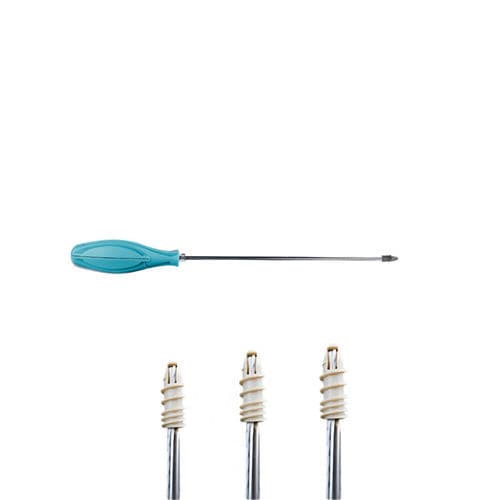
Orthosyn Medikal offers titanium suture anchor options within its arthroscopy portfolio.
Product information:
Orthosyn Titanium Suture Anchors
PEEK Suture Anchors
PEEK stands for polyetheretherketone, a high-performance thermoplastic material used in orthopedic implants.
PEEK anchors provide a non-metallic implant option for selected soft-tissue fixation applications.
Potential characteristics include:
- Non-metallic implant body
- Different anchor diameters
- Compatibility with suture-based fixation
- Material properties distinct from titanium
The use of PEEK does not automatically indicate superior fixation or clinical performance.
Orthosyn Medikal includes PEEK suture anchors within its arthroscopy portfolio.
Product information:
Orthosyn PEEK Suture Anchors
Soft All-Suture Anchors
Soft all-suture anchors use a textile-based fixation concept rather than a conventional rigid screw-type anchor body.
These systems can be inserted through relatively small bone openings, depending on implant design.
Potential considerations include:
- Smaller required bone holes
- Bone preservation
- Textile-based fixation
- Different deployment mechanisms
- Suitability for selected anatomical regions
However, fixation performance depends on bone quality, deployment, implant design, and loading conditions.
Orthosyn Medikal offers soft all-suture anchors in different diameter options within its arthroscopy portfolio.
Product information:
Orthosyn Soft Suture Anchors
PEEK + PEEK Knotless Anchor Systems
PEEK + PEEK knotless anchors use a non-metallic fixation configuration designed for selected arthroscopic soft-tissue fixation procedures.
These systems can provide knotless suture fixation depending on the implant design and surgical technique.
Important considerations include:
- Anchor geometry
- Bone preparation
- Suture tensioning
- Tendon positioning
- Fixation mechanism
- Applicable product indication
Orthosyn Medikal includes PEEK + PEEK knotless fixation technology within its arthroscopy portfolio.
Product information:
Orthosyn PEEK + PEEK Knotless Anchor System
PEEK + Titanium Knotless Anchors
PEEK + Titanium knotless anchors combine different implant materials within a fixation system.
Depending on the design, these systems may support particular insertion and fixation techniques.
Their suitability depends on:
- Bone anatomy
- Implant dimensions
- Surgical approach
- Suture configuration
- Intended use
- Surgeon assessment
Orthosyn Medikal offers PEEK + Titanium knotless anchor technology as part of its arthroscopy portfolio.
Product information:
Orthosyn PEEK + Titanium Knotless Anchors
Why Is Bone Preservation Important in Subscapularis Repair?
Bone preservation is an important consideration in arthroscopic anchor fixation.
Each bone hole removes a certain amount of bone from the implantation site.
The amount of bone removed depends on:
- Anchor diameter
- Implant geometry
- Drilling requirements
- Number of anchors
- Distance between fixation points
- Bone quality
Preserving sufficient bone stock can be relevant when multiple anchors are required or when revision surgery may be necessary.
However, bone preservation must be balanced with the fixation requirements of the repair.
What Is the Role of High-Strength Sutures?
High-strength sutures connect the anchor fixation point to the tendon.
Their purpose is to maintain tendon position against the prepared footprint while biological healing develops.
The mechanical relationship is:
Anchor Fixation → Suture Load Transmission → Tendon Capture → Footprint Contact
Suture selection can influence:
- Tensile characteristics
- Knot behavior
- Tissue interaction
- Suture management
- Repair configuration
However, suture strength alone does not determine the strength of the complete repair.
The construct also depends on anchor fixation, tendon quality, and the suture-tendon interface.
What Is the Relationship Between Subscapularis Tears and Biceps Tendon Instability?
The subscapularis tendon is anatomically associated with structures that stabilize the long head of the biceps tendon.
Injuries involving the superior subscapularis may occur together with disruption of the biceps pulley.
This can contribute to:
- Biceps tendon subluxation
- Biceps tendon dislocation
- Tendon inflammation
- Partial biceps tearing
- Anterior shoulder pain
During arthroscopic assessment, the surgeon may evaluate the biceps tendon and its stabilizing structures.
Depending on the findings, associated biceps pathology may require separate treatment.
Subscapularis repair and biceps treatment are related considerations, but they are not identical procedures.
Can Subscapularis Tears Occur with Supraspinatus Tears?
Yes.
Subscapularis injuries may occur together with supraspinatus tears or more extensive rotator cuff pathology.
Combined tears can influence:
- Tendon mobility
- Repair sequence
- Suture anchor placement
- Number of fixation points
- Tissue tension
- Rehabilitation requirements
In these cases, the surgical plan must consider the entire rotator cuff rather than treating each tendon as an isolated structure.
What Is the Difference Between Partial and Complete Subscapularis Tears?
A partial tear involves only part of the tendon thickness or insertion.
A complete tear involves full-thickness disruption of the affected tendon region.
However, subscapularis tears can also differ in their superior-to-inferior extent.
For example, a full-thickness tear may affect only the upper portion of the tendon rather than its entire width.
This distinction matters because tear size, retraction, and remaining tendon attachment influence repair planning.
Why Is Tendon Quality Important?
Tendon quality affects how sutures interact with the repaired tissue.
Healthy tendon tissue may provide better suture purchase than severely degenerated or fragile tissue.
Factors influencing tendon quality may include:
- Chronic degeneration
- Previous injury
- Tissue thinning
- Fatty infiltration
- Retraction
- Scar formation
Poor tissue quality can increase the risk of suture cut-through or repair failure.
For this reason, surgeons must consider both implant fixation and the condition of the tendon.
Why Is Repair Tension Important?
Appropriate tension is necessary to maintain tendon position without excessively loading the repair.
Excessive tension may place additional stress on:
- Tendon tissue
- Sutures
- Anchors
- Tendon-to-bone interface
Insufficient tension or inadequate reduction may result in poor tendon contact.
The objective is:
Anatomical Reduction + Appropriate Tension + Stable Fixation
Not:
Maximum Suture Tension = Better Repair
What Happens After Arthroscopic Subscapularis Repair?
After surgery, the repaired tendon requires time for biological healing.
Rehabilitation protocols vary according to:
- Tear size
- Repair configuration
- Tissue quality
- Associated procedures
- Surgeon instructions
- Patient characteristics
Postoperative management may include:
- Sling immobilization
- Protected shoulder motion
- Restrictions on active internal rotation
- Avoidance of resisted internal rotation during early healing
- Gradual range-of-motion progression
- Progressive strengthening
- Functional rehabilitation
The exact rehabilitation schedule must follow the treating surgeon's protocol.
How Long Does Subscapularis Tendon Healing Take?
Tendon-to-bone healing is a biological process that develops over time.
Initial mechanical fixation is provided by the repair construct, while biological integration gradually develops at the tendon-bone interface.
The process can be represented as:
Mechanical Fixation → Early Biological Healing → Tendon-to-Bone Integration → Progressive Functional Recovery
Healing and rehabilitation timelines vary significantly.
Factors affecting recovery include:
- Tear size
- Chronicity
- Tendon quality
- Patient age
- Associated injuries
- Repair tension
- Rehabilitation adherence
- Biological healing capacity
Immediate fixation stability should not be confused with complete tendon healing.
Can Arthroscopic Subscapularis Repair Fail?
Yes.
As with other rotator cuff repairs, complications or structural failure can occur.
Potential issues include:
- Tendon retear
- Incomplete tendon healing
- Suture cut-through
- Anchor loosening or pullout
- Loss of fixation
- Persistent weakness
- Shoulder stiffness
- Pain
- Infection
- Recurrent tendon degeneration
The risk depends on the tear, tissue quality, repair technique, patient factors, and postoperative management.
No implant system can completely eliminate these risks.
What Factors Influence Successful Subscapularis Repair?
Successful repair depends on multiple interacting factors.
These include:
- Accurate diagnosis
- Appropriate patient selection
- Complete arthroscopic assessment
- Identification of associated pathology
- Adequate tendon mobilization
- Anatomical footprint preparation
- Correct anchor positioning
- Suitable anchor selection
- Appropriate suture configuration
- Controlled repair tension
- Stable tendon-to-bone contact
- Biological healing
- Postoperative rehabilitation
A useful summary is:
Anatomical Assessment + Tendon Mobility + Footprint Preparation + Stable Fixation + Biological Healing = Foundation of Subscapularis Repair
The quality of the complete repair construct is more important than any single implant characteristic.
What Should Be Considered When Selecting a Suture Anchor?
Parameter Why It Matters
Anchor material Determines implant material characteristics
Anchor diameter Influences bone preparation and available bone stock
Anchor geometry Affects insertion and fixation mechanics
Suture configuration Influences tissue capture and load distribution
Knotted or knotless design Determines the fixation and tensioning approach
Bone quality Influences anchor fixation
Tendon quality Influences suture purchase
Tear dimensions Affect the number and position of fixation points
Footprint anatomy Guides anchor positioning
Applicable indication Determines whether the implant is suitable for the intended procedure
The final selection should follow the relevant product instructions for use and professional surgical assessment.
Orthosyn Suture Anchor Solutions for Arthroscopic Soft-Tissue Repair
Orthosyn Medikal provides a range of suture anchor technologies within its arthroscopy portfolio.
These include:
Orthosyn Titanium Suture Anchors
Metallic fixation options for selected arthroscopic soft-tissue repair procedures.
Orthosyn PEEK Suture Anchors
Non-metallic anchor technology for suture-based fixation.
Orthosyn Soft Suture Anchors
Textile-based fixation options with relatively small bone-hole requirements, depending on the implant.
Orthosyn PEEK + PEEK Knotless Anchors
Knotless fixation technology for selected soft-tissue repair configurations.
Orthosyn PEEK + Titanium Knotless Anchors
Combined-material knotless anchor systems within the Orthosyn arthroscopy portfolio.
These technologies represent different fixation concepts. Their suitability for a particular anatomical site or procedure must be determined according to the applicable product indications, instructions for use, and surgical assessment.
Conclusion
Arthroscopic subscapularis tendon repair is a minimally invasive surgical technique designed to restore the attachment of a torn subscapularis tendon to the lesser tuberosity of the humerus.
The procedure generally involves:
Subscapularis Tear → Arthroscopic Evaluation → Tendon Mobilization → Lesser Tuberosity Preparation → Suture Anchor Placement → Suture Passage → Tendon Fixation → Biological Healing
Suture anchors provide fixation points within the humerus, while high-strength sutures secure the tendon against its anatomical footprint.
Successful repair depends on accurate tear assessment, appropriate tendon mobilization, correct anchor positioning, suitable suture configuration, controlled tensioning, stable tendon-to-bone contact, and postoperative rehabilitation.
Titanium, PEEK, soft all-suture, and knotless anchor systems offer different fixation technologies, but no single implant design is universally appropriate for every subscapularis tear.
Orthosyn Medikal provides titanium, PEEK, soft all-suture, and knotless suture anchor technologies within its arthroscopy portfolio, supporting a range of suture-based soft-tissue fixation strategies. Implant selection for arthroscopic subscapularis tendon repair or other rotator cuff procedures should always be based on the specific product indication, anatomical requirements, tendon and bone quality, and professional surgical judgment.