#Industry News
【Customer Case】Application of Whole Body Plethysmography System in Sleep Research
CeA SST+ Neurons Regulate Stress-Induced Insomnia
Research Overview
Recently, a research team led by Li Chen from the Fujian Key Laboratory of Drug Target Discovery and Structural and Functional Research, Department of Pharmacology, School of Pharmacy, Fujian Medical University, published a study titled "Central amygdala somatostatin neurons modulate stress-induced sleep-onset insomnia" in Communications Biology.
Application of WBP
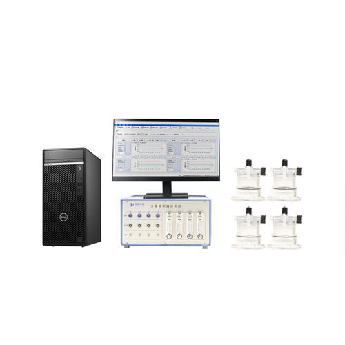
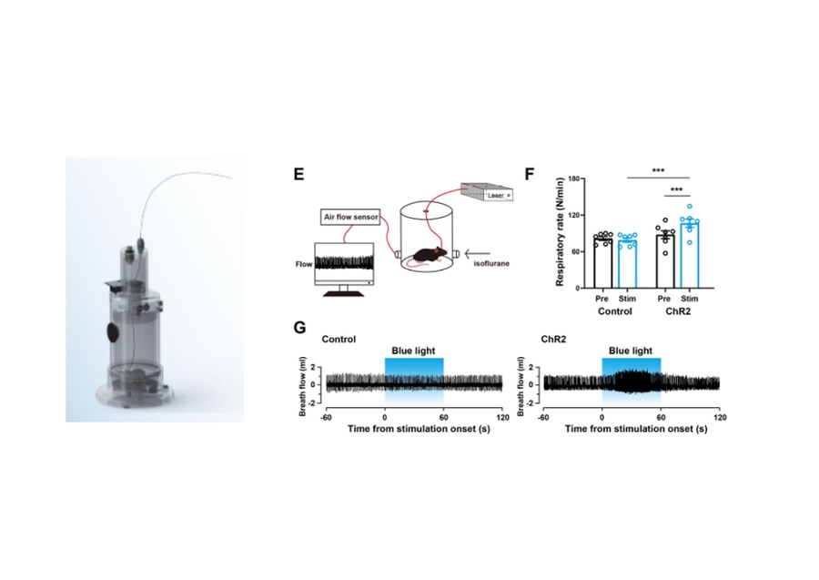
In this study, the team utilized WBP-4M (a whole-body plethysmography system for mice independently developed by TOW-INT TECH) to monitor respiratory parameters. Specifically, under anesthesia, tidal volume (Vt) and respiratory rate were recorded in mice. Blue light stimulation (10 ms, 30 Hz, 3–5 mW/mm², 1 min) was applied to activate somatostatin-positive (SST+) neurons in the central amygdala (CeA) to observe their impact on respiratory frequency.
Results revealed that activating CeA SST+ neurons significantly increased respiratory rate (control: 87.8 ± 6.537 breaths/min → post-stimulation: 106.7 ± 6.926 breaths/min). This indicates that CeA SST+ neurons not only regulate anxiety-like behaviors and sleep-wake transitions but also modulate stress-related physiological metrics (e.g., respiratory rate), further supporting their critical role in stress-induced sleep disorders.
Experimental Methods
1. Fiber Photometry
- Recorded Ca²⁺ dynamics of CeA SST+ neurons under stressors (restraint, rat bedding, air puff, cage change).
- Neurons were labeled using Cre-dependent AAV-GCaMP7s, and fluorescence signals were monitored via optical fibers.
2. Optogenetics
- Expressed ChR2 in CeA SST+ neurons of SST-Cre mice. Acute or semi-chronic activation (20 Hz blue light) was used to assess sleep-wake transitions.
- Combined EEG/EMG recordings analyzed sleep stages (latency and probability of NREM→wake transitions).
3. Chemogenetics
- Employed DREADD technology (hM3Dq activation and hM4Di inhibition) to chronically modulate CeA SST+ neuronal activity, evaluating effects on sleep latency and architecture.
4. Behavioral and Physiological Analyses
- Open Field Test (OFT) & Elevated Plus Maze (EPM): Assessed anxiety-like behaviors.
- WBP-4M: Recorded respiratory rate and tidal volume under optogenetic stimulation.
- Pupillometry: Quantified pupil dilation during CeA SST+ neuron activation.
5. Stress Models
- Cage Change Challenge: Mimicked the "first-night effect" by transferring mice to a cage previously inhabited by other mice.
- Restraint Stress: Restricted mouse movement for 1 hour to induce sleep-onset difficulties.
6. Immunohistochemistry
- Detected c-fos expression to validate neuronal activation and confirmed SST+ neuron specificity via SST antibody labeling.
Experimental Results
1. CeA SST+ Neuronal Responses to Stress
- Multiple stressors (restraint, rat bedding, air puff, cage change) significantly increased Ca²⁺ activity in CeA SST+ neurons (e.g., ΔF/F rose from 4.5% to 17.5% post-cage change).
2. Optogenetic Activation Effects
- Acute activation (20 Hz blue light) shortened NREM→wake latency to 11.6 seconds (vs. control: 60 seconds) with 100% transition probability.
- Semi-chronic activation (1 hour) prolonged sleep latency (35.05 min vs. control: 1.56 min) and increased wake duration (51.19 min vs. 21.20 min).
3. Chemogenetic Modulation
- Activation (CNO treatment) extended sleep latency to 69.6 minutes (vs. control: 18.5 minutes), while inhibition alleviated stress-induced insomnia (latency reduced from 51 to 27 minutes).
- Inhibiting CeA SST+ neurons did not alter physiological sleep architecture, indicating specificity to stress-induced insomnia.
4. Behavioral and Physiological Changes
- CeA SST+ neuron activation induced anxiety-like behaviors (reduced center time in OFT, increased closed-arm time in EPM) and elevated respiratory rate (106.7 vs. baseline: 87.8 breaths/min).
Research Conclusions
By integrating multimodal approaches, the team concluded:
1. CeA SST+ neurons are key regulators of stress-induced sleep-onset insomnia. These neurons are activated under stress, promoting wakefulness and prolonging sleep latency.
2. Target specificity: Inhibiting CeA SST+ neurons alleviated stress-related insomnia without disrupting normal sleep, highlighting their therapeutic potential.
3. Neural mechanism hypothesis: CeA SST+ neurons may regulate sleep-wake balance by suppressing downstream sleep-promoting regions (e.g., vIPAG NTS+ neurons) or integrating upstream stress signals (e.g., PVT, VTA inputs).
Contact us now!
We are committed to making your research easier, more accurate, and more efficient and helping you build confidence in your data! We have provided services for a large number of customers, giving us rich experiences in offering customized, professional solutions according to your needs.
References
[1]Yao W, Huang S X, Zhang L, et al. Central amygdala somatostatin neurons modulate stress-induced sleep-onset insomnia[J]. Communications Biology, 2025, 8(1): 381.