#Industry News
【Nature Sub-journal】Tow-Int's WBP and Laser Speckle Contrast Imaging System Help Decipher Brain Injury Mechanisms
Brain Injury Mechanism Revealed | Tow-Int Tech Supports NC Study
Abstract
Recently, a landmark study led by the Spine Institute of Longhua Hospital, Shanghai University of Traditional Chinese Medicine, has been officially published in the prestigious international journal Nature Communications (IF: 15.7). The research, titled "Brain–cervical lymph node crosstalk contributes to brain injury induced by subarachnoid hemorrhage in mice," was conducted by a team under the guidance of Corresponding Authors Researcher Qianqian Liang and Professor Yongjun Wang, with Assistant Researcher Jinman Chen as the First Author.
This study provides a deep mechanistic explanation for how extravasated erythrocytes in the cerebrospinal fluid following subarachnoid hemorrhage are drained via meningeal lymphatic vessels to cervical lymph nodes, where they are engulfed and degraded by medullary lymphatic endothelial cells, triggering neuroinflammation through the lysosomal protease Cathepsin S. Notably, in this world-class exploratory research, the Laser Speckle Contrast Imaging (LSCI) System and the Whole-Body Plethysmography (WBP) System from Tow-Int Technology provided indispensable technical support for the successful execution and data validation of the study.
Research Background
Subarachnoid hemorrhage (SAH) is a devastating cerebrovascular event with high mortality and disability rates. Its pathological progression stems not only from the initial physical insult of the bleed but is also closely related to subsequent Early Brain Injury (EBI) and Delayed Cerebral Ischemia (DCI). Recent studies have shown that Meningeal Lymphatic Vessels serve as an anatomical connection between the central nervous system (CNS) and the peripheral immune system, draining CNS antigens to Cervical Lymph Nodes (CLNs) and initiating specific immune responses. However, how CLNs participate in the immunopathology of brain injury after SAH, and the precise cellular and molecular mechanisms involved, remained unclear.
Key Findings and Mechanistic Insights
This study systematically dissected the pathway of this immune axis by integrating multiple experimental models and technical approaches:
2.1 Lymph Node Excision Validates Functional Importance:
Researchers surgically removed the mandibular lymph nodes (a major component of the superficial cervical lymph nodes) in mice. They found that in two different SAH models (endovascular perforation and autologous blood injection), the lymph node excision group showed significant improvements in neurological function scores (Modified Garcia scores), reduced brain water content, decreased infiltration of peripheral blood neutrophils (CD11b+Ly6G+), and a lower number of TUNEL+ apoptotic neurons in the cerebral cortex. These results confirmed the promoting role of CLNs in SAH pathology.
2.2 Lymphatic Endothelial Cells (LECs) Mediate Erythrocyte Clearance:
Immunofluorescence staining revealed that from 4 to 24 hours post-SAH, extravasated erythrocytes (TER-119+) co-localized with LYVE-1+ LECs within CLNs, transitioning from intact to degraded morphology over time. In vitro co-culture experiments further confirmed that CFSE-labeled erythrocytes were internalized by LECs. Notably, compared to macrophages (CD169+) in the peripheral areas of the LNs, LECs played a dominant role in the early phase of erythrocyte phagocytosis.
2.3 Single-Cell Sequencing Identifies Key Molecule CTSS:
Single-cell RNA sequencing (scRNA-seq) analysis of CLNs after SAH showed that differentially expressed genes in Medullary LECs were significantly enriched in the Lysosome pathway. Among these, the expression of the gene Ctss, which encodes Cathepsin S (CTSS), was markedly upregulated. Bulk RNA sequencing and qPCR validation in vitro confirmed that co-culture with erythrocytes induced upregulation of Ctss transcripts, along with those of various pro-inflammatory cytokines (e.g., Tnf-α, *Il-1β*, Ccl2), in LECs.
2.4 Genetic and Pharmacological Interventions Validate the Target:
To clarify the function of LEC-derived CTSS, the study constructed a Prox1-creER²; Ctssflox/flox conditional knockout mouse model. After tamoxifen induction, LEC-specific Ctss knockout significantly delayed the degradation rate of extravasated erythrocytes in CLNs and effectively ameliorated SAH-induced neurological deficits and neuroinflammation. Similarly, systemic administration of the small-molecule CTSS inhibitor LY3000328 also produced consistent neuroprotective effects.
Technical Support from Tow-Int's Professional Equipment in Key Experiments
In several critical phases of the mechanistic investigation, the research team relied on Tow-Int's high-precision physiological monitoring equipment to obtain reliable data and rule out potential confounding factors.
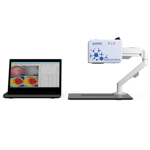
3.1 Application of Tow-Int's Laser Speckle Contrast Imaging (LSCI) System in Cerebral Hemodynamic Analysis:
Scientific Need: To exclude the possibility that the neuroprotective effects of Ctss deletion or pharmacological inhibition were mediated through alterations in Cerebral Blood Flow (CBF) autoregulation, precise quantification of CBF after SAH was required.
Technical Implementation: This study utilized the Tow-Int LSCI system to perform non-invasive, full-field perfusion imaging of the mouse cerebral cortex before SAH induction (baseline), and at 15 minutes and 24 hours post-surgery.
(b) The cerebral blood flow (CBF) detected by the laser speckle blood flow imaging system before SAH, at 15 minutes and 24 hours after SAH surgery in Prox1-CreER T2 /Ctss f/f and Ctss f/f mice.
Data Contribution and Conclusion Support: Data obtained from the system showed that while all groups exhibited the expected sharp decrease in CBF after SAH, there was no statistically significant difference in relative Cerebral Blood Flow (rCBF) at any time point between the Ctss knockout/inhibitor groups and the control groups. This key data robustly demonstrated that the CTSS-mediated neuroprotection was independent of changes in cerebral hemodynamics, thereby focusing the mechanism on the immune regulatory pathway.
3.2 Application of Tow-Int's Whole-Body Plethysmography (WBP) System in Respiratory Function Monitoring:
Synaptic output blockade in SP5C of Nrxn123 cTKO mice significantly reduced NH₃-induced coughing. Chemogenetic inhibition of SP5C neurons strongly suppressed cough responses, while controls remained unaffected.
Scientific Need: In the pharmacological experiments, it was necessary to confirm that the CTSS inhibitor LY3000328 did not suppress basic vital signs, particularly respiratory function, in mice, ensuring that the observed neurological improvements were not a consequence of drug side effects.
Technical Implementation: The study employed the Tow-Int WBP system to perform long-term, non-invasive monitoring of the Respiratory Rate in freely moving mice in a quiet environment.
Data Contribution and Conclusion Support: Respiratory rate data recorded by the WBP system indicated no significant difference between the inhibitor and control groups. This result excluded the possibility that respiratory depression-induced hypoxia confounded the neurological functional assessments, providing crucial physiological evidence for the safety of the CTSS inhibitor and enhancing the rigor of the study's conclusions.
Conclusion
Tow-Int Technology is committed to providing exceptional physiological function research solutions for global scientists, empowering innovative discoveries.
【Reference】
[1] Chen J, Wang J, Zheng W, et al. Brain–cervical lymph node crosstalk contributes to brain injury induced by subarachnoid hemorrhage in mice[J]. Nature Communications, 2025, 16(1): 8551.