#Industry News
【Neuron】Animal Energy Metabolism System Helps Uncover New Mechanism of Energy Expenditure Regulation in the Hypothalamus
[Neuron] Study Pinpoints Key Neurons Controlling Energy Burn, Aided by Animal Metabolism Monitoring System
Abstract
September 17, 2025 – A research paper titled "Identification of a neural basis for energy expenditure in the mouse arcuate hypothalamus" was published online in Neuron (IF=15) by corresponding authors Qing-Feng Wu from the Institute of Genetics and Developmental Biology, Chinese Academy of Sciences, and Peng Cao from the National Institute of Biological Sciences. This study identified a hypothalamic GABAergic neuronal subtype, characterized by Crabp1 expression, which targets multiple nuclei to regulate energy expenditure in mice.
This research represents a breakthrough in the field of neurometabolism and establishes a new technical and methodological paradigm for energy metabolism studies. It is noteworthy that the study utilized an Animal Energy Metabolism Monitoring System for phenotypic analysis of energy metabolism, accurately capturing the dynamic process of neuronal regulation of energy expenditure and providing robust data support for the conclusions.
Research Background
Obesity has become a global health issue. Traditional weight-loss strategies primarily focus on controlling energy intake, such as dieting or pharmacological appetite suppression. However, these methods are often accompanied by the challenge of weight rebound. In recent years, increasing energy expenditure has been regarded as a more sustainable intervention strategy for obesity. Yet, how the brain regulates energy expenditure and the specific neural circuit basis remains unclear.
The hypothalamic arcuate nucleus (ARC) is a central hub for energy balance regulation. AgRP and POMC neurons serve as the classic model for "feeding-energy expenditure" regulation, promoting feeding/inhibiting energy expenditure and inhibiting feeding/promoting energy expenditure, respectively. However, the ARC also contains a large number of "non-AgRP, non-POMC" neurons, whose functions and mechanisms remain poorly defined.
Research Design and Key Findings
1. Discovery and Identification of Crabp1 Neurons
Using single-cell and single-nucleus RNA sequencing technologies, the research team identified for the first time a GABAergic neuronal subtype in the ARC expressing the Crabp1 gene. These neurons do not express various known neuroendocrine markers such as AgRP, POMC, GHRH, or kisspeptin, indicating unique molecular characteristics and spatial distribution.
Further research revealed that Crabp1 neurons exhibit low expression of leptin and insulin receptors but high expression of various neurotransmitter receptors (e.g., glutamate, 5-HT, GABA receptors), suggesting their activity is primarily regulated by neural circuits rather than circulating hormones.
Figure 1. Identification of Crabp1 neurons as a population of “non-AgRP, non-POMC” neurons in the ARC
2. Functional Validation: Crabp1 Neurons as the "Central Switch" for Energy Expenditure
To clarify the function of Crabp1 neurons, researchers employed genetic tools for precise manipulation:
Inhibiting Crabp1 neurons: Mice exhibited reduced spontaneous physical activity, decreased body temperature, diminished cold-induced thermogenesis, ultimately leading to weight gain and fat accumulation.
Figure 2. Synaptic inactivation of Crabp1 neurons suppresses energy expenditure and causes obesity
Activating Crabp1 neurons: Significantly increased energy expenditure, oxygen consumption, locomotor activity, and brown adipose tissue thermogenesis, even conferring some resistance to high-fat diet-induced obesity.
Figure 3. Chemogenetic activation of Crabp1 neurons promotes energy expenditure
Notably, Crabp1 neurons simultaneously regulate both energy expenditure and food intake. Their mode of action was summarized by the authors as the "mirror imbalanced model", forming a sharp contrast to the traditional AgRP/POMC "seesaw model".
3. Neural Dynamics: Cold and Exercise Activate Crabp1 Neurons
Using cFos staining and in vivo fiber photometry calcium recording, the study found that Crabp1 neurons are specifically activated during cold exposure and exercise, but show no significant response to hunger, satiety, or restraint stress. This indicates that these neurons primarily respond to physiological and environmental stimuli related to energy demand, rather than energy status per se.
Figure 4. Crabp1 neurons respond to cold exposure and physical activity
4. Circuit Mechanism: "One-to-Many" Projection Pattern Coordinately Regulates Multi-dimensional Energy Expenditure
Using whole-brain scale single-neuron tracing techniques, researchers found that Crabp1 neurons primarily project to multiple hypothalamic nuclei (e.g., MPOA, PVN, LH, DMH) and basal forebrain regions (e.g., BNST). Particularly noteworthy, Group 2 neurons innervate multiple target regions simultaneously via axonal collateral branches, forming a "one-to-many" circuit architecture that enables coordinated control over multi-dimensional aspects of energy expenditure, including locomotion, thermogenesis, and body temperature.
Figure 5. Mapping the input and output connectivity of Crabp1 neurons
Figure 6. Crabp1 neurons coordinate energy expenditure via axon collateral branches
5. Intraperitoneal Injection of AOSs Was Also Effective, Suggesting Systemic Action
Prolonged light exposure (18 hours light / 6 hours dark) suppressed the spontaneous firing of Crabp1 neurons, reduced energy expenditure, and led to weight gain. Chemogenetic reactivation of these neurons reversed the metabolic decline induced by the long photoperiod. This finding provides a potential neural mechanism underlying the link between light pollution and metabolic diseases.
Figure 7. Susceptibility of Crabp1 neurons to long photoperiods and their activation in mitigating diet-induced obesity
Technical Highlight: The Precision Tool for Energy Metabolic Phenotyping
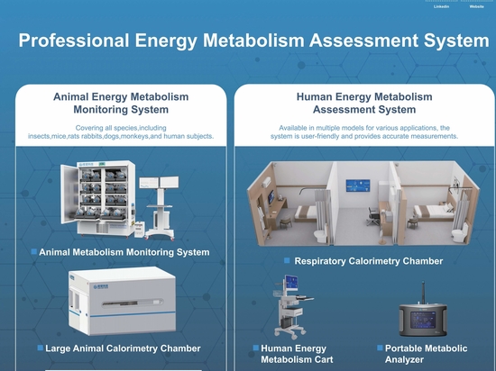
In this study, the continuous monitoring of key metabolic parameters – including energy expenditure, oxygen consumption, carbon dioxide production, respiratory exchange ratio (RER), and locomotor activity – relied on the high-precision Animal Energy Metabolism Monitoring System. This system enables:
Real-time, synchronous monitoring of multiple metabolic parameters, avoiding errors from data desynchronization.
Long-term, continuous recording, capturing dynamic changes under circadian rhythms and environmental interventions.
High-throughput parallel experiments, supporting large sample sizes and controlled conditions.
Behavioral analysis compatibility, simultaneously recording voluntary activity and metabolic indices.
If you are interested in this system or wish to learn more about its application cases, please feel free to contact the tow-int team. We are committed to providing you with professional technical support and customized solutions.
TOW-INT Animal Energy Metabolism Monitoring System – Providing Visible Data Power for Energy Metabolism Research.
【Reference】
[1] Wang T, Han S, Wang Y, et al. Identification of a neural basis for energy expenditure in the mouse arcuate hypothalamus[J]. Neuron, 2025.