#Industry News
Application of Energy Metabolism Monitoring Systems in Fatty Liver Mouse Models
Metabolism System can be used in mouse models of fatty liver.
Fatty Liver Disease and Its Association with Obesity
Fatty liver is closely associated with obesity and is often accompanied by metabolic disorders such as insulin resistance and dyslipidemia. Obesity, particularly visceral obesity, is one of the main risk factors for Metabolic Dysfunction-Associated Fatty Liver Disease (MAFLD). Therefore, establishing appropriate animal models of fatty liver is essential for in-depth studies on the pathogenesis of MAFLD and for exploring potential therapeutic strategies.
Energy Metabolism Differences in Fatty Liver Mouse Models
Mouse models of fatty liver (typically induced by high-fat diets), obese mouse models, and normal control mice exhibit significant differences in energy metabolism. These differences are primarily reflected in oxygen consumption (VO₂), carbon dioxide production (VCO₂), respiratory exchange ratio (RER), energy expenditure (EE), and activity levels. The following sections detail each of these parameters:
1. Oxygen Consumption (VO₂) and Carbon Dioxide Production (VCO₂)
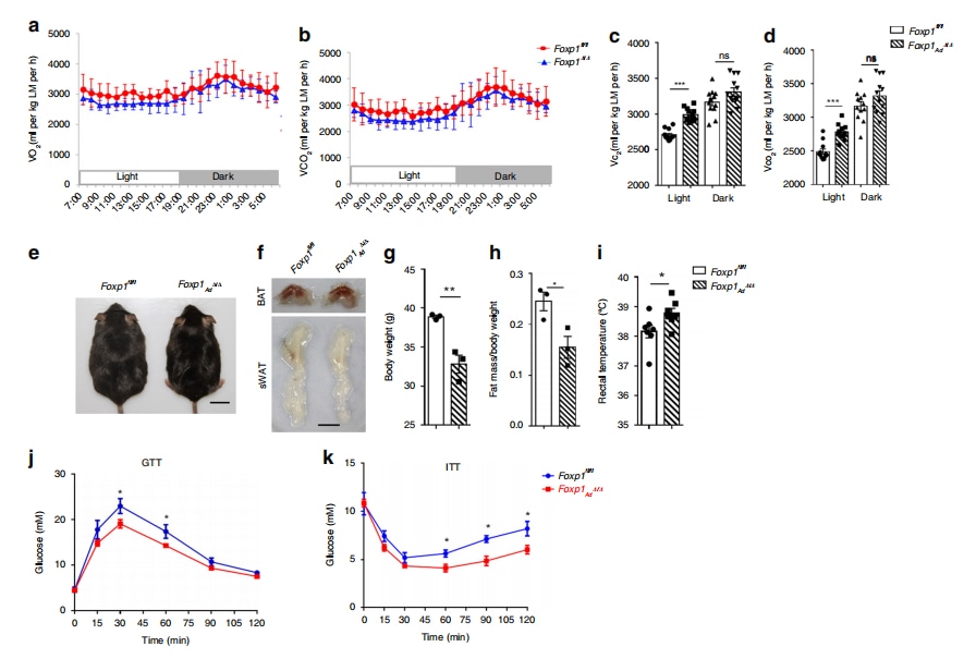
Fatty liver/obese model mice vs. control group: Fatty liver and obese model mice usually exhibit lower VO₂ and VCO₂. For example, one study found that deletion of the Foxp1 gene led to increased activity of brown adipose tissue, elevated energy expenditure, and resistance to high-fat diet-induced obesity (Liu et al., 2019). As shown in Figure 1, Foxp1 knockout mice (Foxp1⁻/⁻) had higher oxygen consumption than Foxp1⁺/⁺ mice (Liu et al., 2019).
Long-term high-fat feeding impairs mitochondrial function and energy metabolism, reducing oxygen consumption. Studies show that high-fat diets alter respiratory function and affect circadian rhythms in mice (Wu et al., 2024).
Spexin modulates energy metabolism by increasing oxygen consumption. One study demonstrated that Spexin enhances browning of white adipose tissue via the JAK2-STAT3 pathway, improving obesity-related metabolic disorders (Zeng et al., 2024).
2. Respiratory Exchange Ratio (RER)
Definition:
RER is the ratio of carbon dioxide production to oxygen consumption and reflects the type of substrate being metabolized. An RER close to 1 indicates carbohydrate utilization, whereas an RER near 0.7 suggests fat utilization.
Fatty liver/obese model mice vs. control group: These mice typically have a lower RER, indicating a greater reliance on fat as an energy source (Liu et al., 2019). In the same study, Figure 1 shows that RER values in Foxp1-deficient mice were lower than in controls, suggesting enhanced lipid metabolism due to Foxp1 deletion.
3. Energy Expenditure (EE)
Fatty liver/obese model mice vs. control group: Although these models have lower RERs, their EE is not necessarily reduced and depends on several factors including activity level, food intake, and metabolic adaptation.
Effect of temperature:
Environmental temperature significantly influences EE. Studies show that mice consume more energy to maintain body temperature at standard room temperature (John et al., 2022).
4. Activity Level
Fatty liver/obese model mice vs. control group: Fatty liver and obese mice generally have lower activity levels, possibly due to weight gain, insulin resistance, and inflammation.
Voluntary exercise can modulate hepatic immune phenotypes and improve metabolic parameters (Gehrke et al., 2019).
5. Summary
In summary, fatty liver and obese mouse models generally show lower oxygen consumption, lower RER, variable energy expenditure, and reduced physical activity. However, these indices are influenced by multiple factors such as diet composition, genetic background, environmental temperature, and experimental conditions. For example, in C57BL/6J mice, the duration of dietary intervention is crucial. Marvyn et al. (2016) reported that short-term (3-day) high-fat feeding increases fat oxidation, while long-term feeding leads to different metabolic outcomes.
Hence, when comparing energy metabolism among different groups, it is important to consider all these variables and apply rigorous statistical analysis.
Broader Significance of Metabolism System
Besides elucidating the metabolic characteristics of fatty liver, energy metabolism systems in animals are valuable in the following ways:
Assessing the Progression of Fatty Liver
Early Detection: Metabolic alterations often precede histological liver changes. Monitoring energy metabolism may enable early detection and timely intervention.
Disease Monitoring: Continuous tracking of metabolic parameters can reveal disease progression or improvement, helping guide therapeutic adjustments.
Understanding Pathogenesis
Glucose and Lipid Metabolism Disorders: By analyzing metabolic data, researchers can investigate the roles of insulin resistance, lipid synthesis, and lipolysis in fatty liver pathogenesis.
Oxidative Stress and Inflammation: Fatty liver often involves oxidative stress and inflammation. Combining metabolic with other biochemical data can help clarify their interrelationships.
Evaluating Therapeutic Strategies
Drug Efficacy: Metabolic systems can evaluate how drugs affect energy metabolism in animal models. For instance, changes in RER and EE can indicate whether a drug improves metabolic function.
Nutritional Interventions: Studies on different diets (e.g., high-fat or low-carb) and their metabolic effects can inform evidence-based nutritional strategies.
Predicting Disease Risk and Prognosis
Risk Assessment: Metabolic markers such as elevated RER may signal increased fatty liver risk, aiding in early identification of at-risk individuals.
Prognostic Evaluation: Persistent metabolic disturbances may predict disease progression and poor outcomes.
Synchronized data recording and analysis to ensure data accuracy and safety
Advancing Basic and Translational Research
Mechanistic Insights: Animal studies of energy metabolism deepen understanding of fatty liver pathology and support clinical hypothesis generation.
Translational Applications: Findings from animal models can be integrated into clinical research, accelerating the development of new diagnostic tools and therapies.
Ethical and safety compliance ensures welfare for both animals and human participants
Animal Energy Metabolism Monitoring System
One of the fundamental characteristics of life is active energy metabolism—the constant intake and consumption of energy. In mammals, most energy comes from the oxidation of three macronutrients: carbohydrates, fats, and proteins.
The Animal Energy Metabolism Monitoring System independently developed by Tawang Technology estimates energy production by analyzing oxygen–carbon dioxide exchange during metabolism. By evaluating oxygen consumption (O₂) and carbon dioxide production (CO₂), the system determines the energy content of consumed food. It also supports monitoring of multiple parameters and the integration of various optional features.
References:
[1]Liu P, Huang S, Ling S, et al. Foxp1 controls brown/beige adipocyte differentiation and thermogenesis through regulating β3-AR desensitization[J]. Nature communications, 2019, 10(1): 5070.[2] Zeng B, Shen Q, Wang B, et al. Spexin ameliorated obesity-related metabolic disorders through promoting white adipose browning mediated by JAK2-STAT3 pathway[J]. Nutrition & Metabolism, 2024, 21(1): 22.
[3] Gehrke N, Biedenbach J, Huber Y, et al. Voluntary exercise in mice fed an obesogenic diet alters the hepatic immune phenotype and improves metabolic parameters–an animal model of life style intervention in NAFLD[J]. Scientific reports, 2019, 9(1): 4007.
[4] Marvyn P M, Bradley R M, Mardian E B, et al. Data on oxygen consumption rate, respiratory exchange ratio, and movement in C57BL/6J female mice on the third day of consuming a high-fat diet[J]. Data in brief, 2016, 7: 472-475.
[5]John L M, Petersen N, Gerstenberg M K, et al. Housing-temperature reveals energy intake counter-balances energy expenditure in normal-weight, but not diet-induced obese, male mice[J]. Communications biology, 2022, 5(1): 946.
[6]Wu Y, Yang M, Wu S, et al. Zinc finger BED-type containing 3 promotes hepatic steatosis by interacting with polypyrimidine tract-binding protein 1[J]. Diabetologia, 2024, 67(10): 2346-2366.