#Industry News
Using Whole-Body Plethysmography to Study and Monitor Cough Mechanisms in Mice
Whole-Body Plethysmography for Mouse Cough Behavior Research
Abstract
Coughing is an essential respiratory defense mechanism that helps clear mucus or foreign substances from the airways. Under normal conditions, it is a powerful and effective physiological response. However, in pathological states, the neural mechanisms controlling these defensive behaviors can become hypersensitive, leading to chronic dry coughing that severely impacts quality of life. Although cough is a common reason for patients seeking medical help, current treatments remain limited and may lead to drug abuse, largely due to an insufficient understanding of the neural and molecular mechanisms underlying cough behavior.
A groundbreaking study led by Professor Luo Fujun’s team at Guangzhou Laboratory, published in eLife, identified a brainstem circuit that controls cough-like airway defensive behaviors in mice. The first author, Professor Xu Xiaoshan, utilized a freely moving mouse model to reveal the brainstem neural circuit mediating evoked respiratory reflexes, defining it as a cough-like response. By employing multiple neural circuit mapping and modulation techniques, the team discovered a previously unrecognized role of the caudal spinal trigeminal nucleus (SP5C) in reflexive cough behavior.
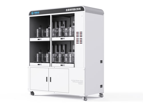
This study combined whole-body plethysmography (WBP), audio recording, and video tracking technologies developed by Tow-Int Tech to establish a quantitative paradigm for studying cough behavior in awake mice. Using TRAP2 transgenic mice and in vivo fiber photometry, the researchers demonstrated that neural activity in SP5C is highly correlated with cough responses induced by tussive agents.
Further experiments showed that inhibiting synaptic output from SP5C or suppressing its activity via chemogenetic methods effectively eliminated these cough reflexes. Conversely, optogenetic stimulation of SP5C excitatory neurons or their projections to the ventral respiratory group (VRG) triggered robust cough-like behaviors even in the absence of tussive stimuli.
Notably, sustained enhancement of SP5C neuronal excitability led to chronic spontaneous coughing in mice. These findings provide strong evidence for the existence of a previously unidentified brainstem circuit (SP5C→VRG) that controls cough defense behaviors in mice.
Research Objectives
This study systematically identified and validated the central neural circuit controlling cough behavior using a mouse model and various neural circuit analysis technologies, with a particular focus on the role of SP5C.
Materials and Methods
A multidisciplinary approach was employed to analyze cough behavior from behavioral, neural activity, circuit connectivity, and functional modulation perspectives.
3.1 Animal Models
Wild-type (WT), TRAP2 transgenic, neurexin 1/2/3 conditional knockout (Nrxn123 cTKO), VGluT2-IRES-Cre, and GAD2-IRES-Cre transgenic mice were used for cell-type-specific manipulations.
3.2 Cough Induction and Monitoring
Tussive stimuli: Citric acid (CA), capsaicin, and ammonia (NH₃) were nebulized to induce cough-like responses.
Monitoring system: Whole-body plethysmography (WBP) recorded respiratory airflow changes, while microphones and cameras captured cough sounds and movements for multimodal quantitative analysis.
Cough definition: A cough-like event was defined by the characteristic "inspiration-compression-expiration" triphasic airflow pattern in WBP, accompanied by distinct cough sounds.
3.3 Neural Activity Monitoring
In vivo fiber photometry: AAV-GCaMP6s was injected into target brain regions (e.g., SP5C, NTS, VRG) to monitor calcium signals and assess temporal correlations with cough behavior.
3.4 Necessity Validation
Synaptic output blockade: AAV-Cre was injected into SP5C of Nrxn123 cTKO mice to conditionally block synaptic transmission.
Chemogenetic inhibition: AAV-hM4Di-mCherry was injected into SP5C, and deschloroclozapine (DCZ) was administered intraperitoneally to inhibit neuronal activity.
3.5 Sufficiency Validation
Optogenetic activation: AAV-ChrimsonR was injected into SP5C to stimulate excitatory neurons (CaMKII+). Projection-specific activation was achieved by injecting AAV-Cre into SP5C and AAV-DIO-ChrimsonR into VRG.
3.6 Circuit Tracing
Anterograde and retrograde trans-synaptic tracing:
Anterograde: AAV-hSyn-EGFP was injected into SP5C to visualize axonal projections.
Retrograde: Pseudorabies virus (RV) was injected into VRG to label monosynaptic input neurons from SP5C.
Dual-virus strategy: AAV2/1-Cre (anterograde) and AAV2/9-DIO-Chrimson-mCherry were used to confirm direct synaptic connections between SP5C and VRG.
3.7 Chronic Cough Model
NaChBac, a bacterial voltage-gated sodium channel, was overexpressed in SP5C to enhance neuronal excitability and induce spontaneous coughing.
3.8 Electrophysiological Recording
Whole-cell patch-clamp recordings were performed on acute brainstem slices to examine SP5C neuronal properties (e.g., action potential firing, membrane resistance).
Results
4.1 Establishment and Validation of Cough Behavior Model
NH₃, CA, and capsaicin induced typical cough-like responses characterized by the triphasic airflow pattern and distinct cough sounds. NH₃ was selected as the primary tussive stimulus due to its strong and consistent effects.
Control (saline) treatments produced negligible coughing, confirming model specificity.
4.2 SP5C Neural Activity Is Highly Correlated with Cough
TRAP2 labeling showed that NH₃ stimulation activated neurons in SP5C, NTS, and VRG. Fiber photometry confirmed that SP5C calcium signals significantly increased during cough events and were synchronized with cough timing.
4.3 SP5C Is Necessary for Cough Reflex
Synaptic output blockade in SP5C of Nrxn123 cTKO mice significantly reduced NH₃-induced coughing. Chemogenetic inhibition of SP5C neurons strongly suppressed cough responses, while controls remained unaffected.
4.4 Activation of SP5C Directly Triggers Cough
Optogenetic activation of VGluT2+ or GAD2+ neurons only altered respiratory rhythm without inducing coughing. In contrast, stimulating CaMKII+ excitatory neurons in SP5C triggered robust cough-like behaviors without tussive stimuli.
4.5 SP5C Regulates Cough via Direct Projections to VRG
Tracing experiments revealed direct projections from SP5C to cVRG, rVRG, and preBötC. Retrograde viral tracing confirmed monosynaptic connections between SP5C and VRG. Optogenetic activation of SP5C→VRG projections induced coughing, with high-frequency stimulation being more effective.
4.6 Enhanced SP5C Excitability Induces Chronic Spontaneous Cough
NaChBac overexpression in SP5C led to spontaneous coughing within 3–4 days, with frequency increasing over time. These mice also exhibited enhanced sensitivity to NH₃, with stronger and faster cough responses. Electrophysiological recordings confirmed increased excitability and spontaneous action potentials in SP5C neurons.
Conclusion
This study systematically identified and validated the central neural circuit controlling cough behavior in mice, highlighting the critical role of SP5C.
Key findings include:
- **SP5C is a key central nucleus for cough reflex**: Its neural activity is synchronized with coughing, its inhibition eliminates cough, and its activation triggers cough.
- **SP5C directly regulates VRG via monosynaptic connections**: This pathway forms the neural basis for cough motor output.
- **SP5C hyperexcitability causes chronic coughing**: Enhanced SP5C neuronal excitability induces spontaneous coughing and cough hypersensitivity, providing insights into chronic cough mechanisms.
- **Mice are a valid model for studying cough neural mechanisms**: Multimodal technologies confirmed the reliability of mouse cough-like behavior, supporting the use of mice in neural circuit research.
**Significance and Outlook**
This study reveals a previously overlooked cough regulatory center (SP5C) and offers new mechanistic insights into chronic cough and cough hypersensitivity. It may pave the way for developing centrally targeted antitussive therapies.
【Reference】
Xu X, Nie X, Zhang W, et al. A brainstem circuit controls cough-like airway defensive behaviors in mice. *bioRxiv*, 2024: 2024.09.08.611924.