#Product Trends
Pulmonary Vein Cryoablation: Essential Knowledge for Cardiac Intervention Training
Pulmonary Vein Cryoablation: Essential Knowledge for Cardiac Intervention Training
Atrial fibrillation (AF) is one of the most common cardiac arrhythmias worldwide, and pulmonary vein isolation (PVI) has become a cornerstone in its interventional treatment. Among the available technologies, pulmonary vein cryoablation has gained significant recognition for its safety, efficiency, and reproducible therapeutic outcomes. As clinical demand grows, high-fidelity anatomical models play a crucial role in helping physicians master the complex steps of catheter-based ablation procedures.
What Is Pulmonary Vein Cryoablation?
Pulmonary vein cryoablation is a minimally invasive procedure designed to electrically isolate the pulmonary veins from the left atrium. In many patients with AF, abnormal electrical impulses originate within the pulmonary veins. By creating a continuous ring-shaped lesion around each vein’s ostium, cryoablation prevents these triggers from entering the atrium and initiating arrhythmias.
Unlike radiofrequency ablation—which uses high-frequency electrical energy to heat tissue—cryoablation works by delivering extremely low temperatures through a balloon catheter. When the balloon is inflated and positioned at the pulmonary vein opening, controlled freeze cycles form a stable lesion without excessive tissue damage. This “cryo-adhesion” also helps keep the balloon securely in place during freezing, contributing to more predictable outcomes and easier catheter manipulation.
How the Procedure Is Performed
1.Vascular Access
Most procedures begin with femoral venous access, though internal jugular access may be used in specific situations.
2.Atrial Septal Puncture
The catheter is advanced through the inferior vena cava and guided into the right atrium. A transseptal puncture provides access to the left atrium, a crucial step that requires precision and extensive practice.
3.Cryoballoon Positioning
Once inside the left atrium, the cryoballoon is navigated to each pulmonary vein ostium. Proper positioning is essential for achieving full circumferential contact.
4.Freezing Cycles
Freeze–thaw cycles are applied to create durable lesions. Temperature, balloon stability, occlusion quality, and real-time monitoring of adjacent structures are key to procedural success.
Cryoablation’s streamlined workflow and shorter learning curve make it particularly attractive in clinical practice—yet mastery still requires guided simulation and repeated hands-on experience.
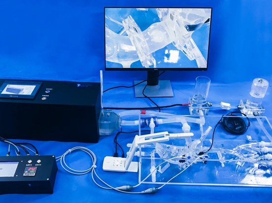
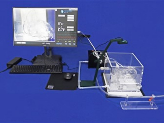
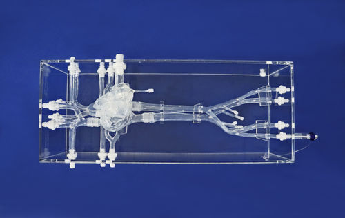
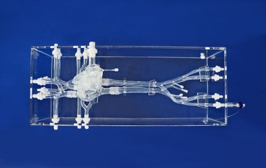
In training and device development, realistic anatomical simulation is essential. Our arteriovenous model—extending from femoral access points to the complete pulmonary arterial and venous system—provides an ideal platform for practicing key steps of pulmonary vein cryoablation. Trainees can simulate catheter navigation from the femoral veins and internal jugular vein, perform atrial septal puncture, and rehearse cryoballoon positioning within anatomically accurate pulmonary veins. The modular heart design and transparent acrylic structure also support product demonstrations, device testing, and educational instruction for cardiac intervention technologies.
By replicating complex vascular pathways and heart anatomy, this model enables safe, repeatable, and highly realistic training for pulmonary vein cryoablation as well as other interventional procedures critical to modern arrhythmia management.