#Product Trends
Understanding Atrial Septal Defect (ASD)
Understanding Atrial Septal Defect (ASD)
The heart is a powerful organ that pumps blood throughout the body, delivering oxygen and nutrients to tissues and removing waste products. It has four chambers — two upper chambers called the atria and two lower chambers called the ventricles. Normally, the left and right atria are separated by a wall called the atrial septum.
An Atrial Septal Defect (ASD) is a congenital condition where there is a hole in this wall, allowing blood to flow between the left and right atria. This abnormal opening can vary in size and may close on its own during infancy, or persist into adulthood if left untreated.
What Happens in ASD?
In a healthy heart, oxygen-rich blood from the lungs returns to the left atrium and is pumped out to the rest of the body. In individuals with ASD, some of this oxygen-rich blood leaks through the hole into the right atrium, mixing with oxygen-poor blood. This results in an increased volume of blood flowing to the lungs, which can overwork the heart and lead to complications such as:
Shortness of breath
Fatigue
Heart palpitations
Increased risk of stroke
Pulmonary hypertension (high blood pressure in the lungs)
Diagnosis and Treatment
ASD is often diagnosed using imaging techniques like echocardiography, CT scans, or MRI. Small defects might not cause symptoms and may not need treatment. Larger defects, however, often require closure through surgery or catheter-based procedures using specialized devices.
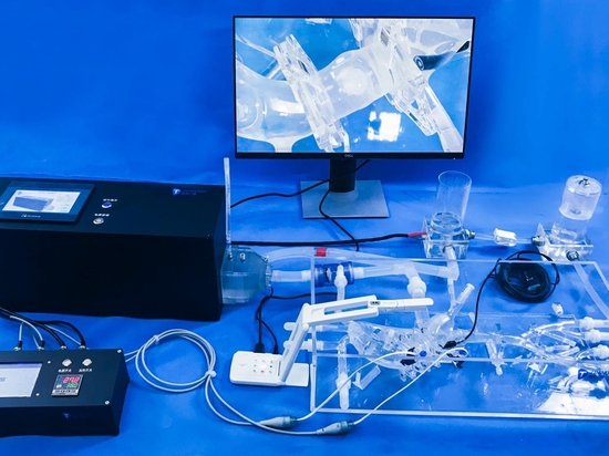
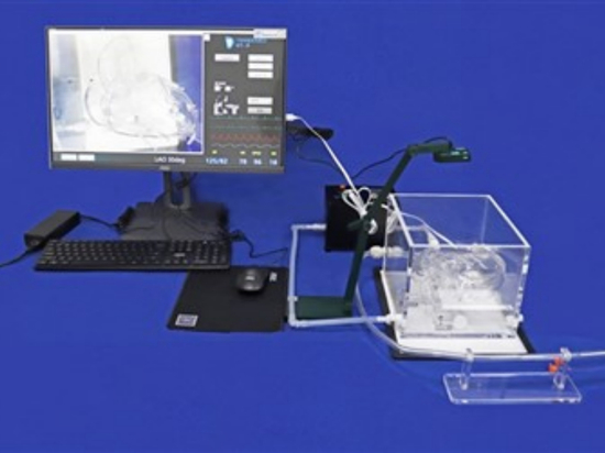
Our ASD Medical Simulation Model
To assist in the training of medical professionals and support research in cardiac interventions, we have developed a realistic ASD simulation model. This model provides a detailed and hands-on experience for practicing various interventional procedures.
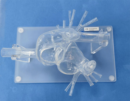
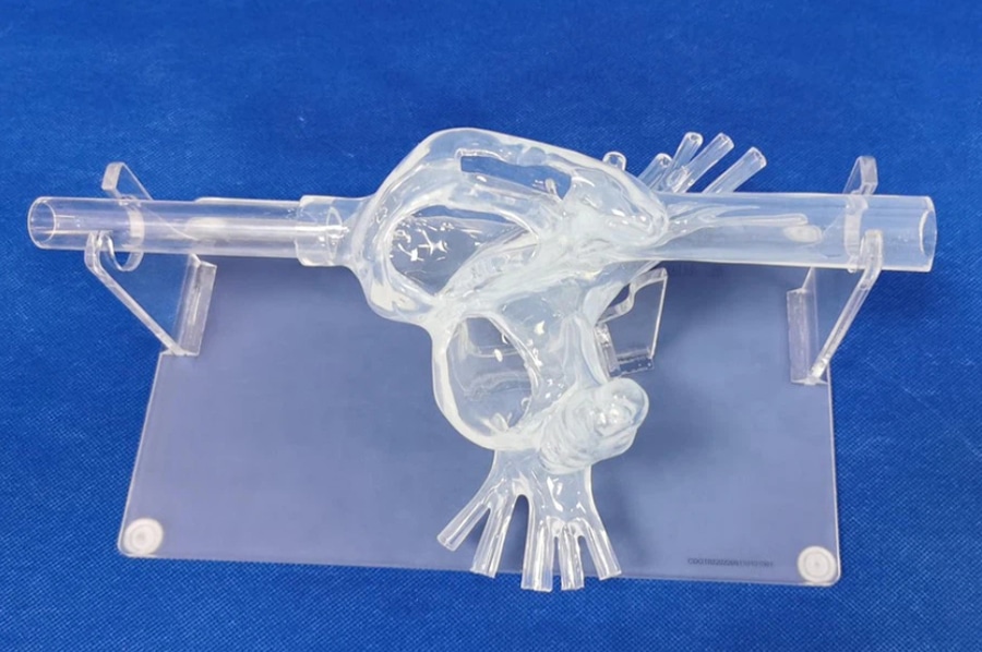
Model Features:
Anatomical Structures Included: Left & right atrium, pulmonary vein, superior vena cava (SVC), inferior vena cava (IVC), left atrial appendage (LLA), right atrial appendage (RAA), and foramen ovale.
Transparent Design: The model is mounted on a clear acrylic plate, allowing full visibility of internal structures.
IVC Extension: The IVC is extended using a transparent resin tube to better simulate catheter access.