#Industry News
How Simulated Anatomy Solves Five Key Pain Points in Aortic Valve Development with TAVR Training Simulators
How Simulated Anatomy Solves Five Key Pain Points in Aortic Valve Development with TAVR Training Simulators
Overview: Core Challenges in Aortic Valve R&D
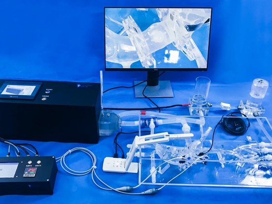
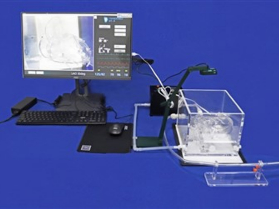
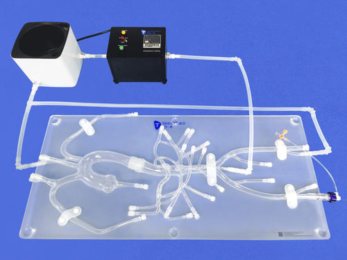
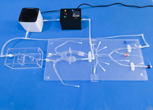
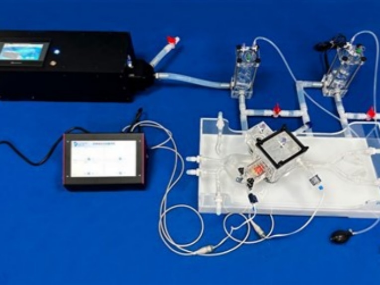
The development of aortic valve systems demands a balanced focus on work efficiency, R&D costs and regulatory compliance. Before clinical trials kick off, nearly all R&D teams encounter common technical challenges during product iteration. Many developers also rely on professional TAVR training simulators for preliminary verification and technical demonstration. Typical doubts include: Will the opening and closing movements of valve leaflets cause premature damage? Is the delivery system flexible enough to pass through the aortic arch smoothly? What is the actual risk of paravalvular leakage under severe and complex calcified lesions? Traditional static testing fails to replicate the real physiological environment of the human heart. Fortunately, customized 3D-printed aortic valve anatomical models and supporting TAVR training simulators from Trandomed effectively address all the above pain points for medical device R&D.
1. Rectify Distorted Hemodynamic Data
First, it eliminates distorted hemodynamic data. Ordinary basic models cannot simulate vascular elasticity and pressure variations at different vascular segments. Our customized valve models, paired with proprietary hemodynamic systems, generate highly realistic and adjustable pressure waveforms. Researchers can measure transvalvular pressure gradient, effective orifice area (EOA) and regurgitant flow in real time. These parameters are highly consistent with in-vivo conditions, delivering solid and reliable data to support the development of prosthetic heart valves.
2. Reduce Hidden Costs in Leaflet Design
Second, it cuts down hidden costs in leaflet design. Problems such as insufficient leaflet opening or asynchronous closure are usually detected only after long-term tests, leading to extra hidden expenses. Our specially made high-transparency models, combined with high-speed cameras, allow teams to observe the kinematics of valve leaflets clearly. Engineers can quickly locate stress concentration areas and optimize leaflet cutting solutions, greatly saving time and overall R&D investment.
3. Resolve Passability Issues of Delivery Systems
Third, it tackles the navigational challenges of delivery systems. Standard testing setups cannot restore the resistance of complex aortic arch anatomies during device delivery. Our aortic models are 1:1 reconstructed based on real patient CT and angiography data, featuring authentic anatomical bends. Connected to a pulsatile system, they realistically simulate the whole process of valve crossing, deployment and retrieval, enabling accurate evaluation of delivery system flexibility and valve expansion performance.
4. Early Identification of Paravalvular Leakage (PVL)
Fourth, it enables early detection of paravalvular leakage (PVL). Uniform valve ring models lack the realism to reproduce PVL, a major clinical concern. We provide models with mild, moderate and severe calcification to restore irregular valve ring structures. Developers can test the sealing performance of valve skirts against calcified gaps in the lab and optimize anti-leakage designs in advance.
5. Improve Practical Experience for Training & Demonstration
Fifth, it enhances practical experience for demonstration and training. Theoretical lectures and 2D images cannot help doctors perceive the physical feedback of medical devices intuitively. Our 3D-printed heart valve models, integrated with hemodynamic systems and professional TAVR training simulators, fully mimic human physiological conditions. They support accurate functional tests, advanced clinical training and product demonstrations, shortening doctors’ learning curves and building their recognition of your products.
Product Specifications
Data Source: 1:1 optimized design based on real human anatomical data
Lesion Types: Healthy valves, valvular insufficiency, mild/moderate/severe calcification
Functional Compatibility: Compatible with hemodynamic systems for dynamic feedback
Application Scenarios: Pre-market testing, clinical training and product demonstration
Conclusion: Accelerate R&D from Lab to Clinic
Trandomed’s customized medical models and complete TAVR training simulators deliver reliable in-vitro simulation solutions for the early stage of medical device development. They effectively accelerate the transition from laboratory research to clinical application. Feel free to contact our technical consultants to get detailed configuration plans and professional support.