#Product Trends
Five Major Pain Points in Aortic Valve Development: How to Overcome Them Through Anatomical Simulation?
Five Major Pain Points in Aortic Valve Development: How to Overcome Them Through Anatomical Simulation?
The development of the aortic valve system requires balancing efficiency, R&D costs, and regulatory compliance. Before entering clinical trials, every R&D team faces similar questions:
• "Will the opening and closing motion of the valve leaflets lead to early rupture?"
• "Is the delivery system sufficiently smooth when passing through the aortic arch?"
• "How great is the risk of paravalvular leakage in complex calcified lesions?" Traditional static testing struggles to simulate the real physiological environment of the heart, but the innovative 3D-printed aortic valve anatomical model effectively addresses these R&D pain points.
I. Solving the Problem of Hemodynamic Data Distortion
Conventional, simplified models cannot simulate real vascular elasticity and pressure data at different vascular locations.
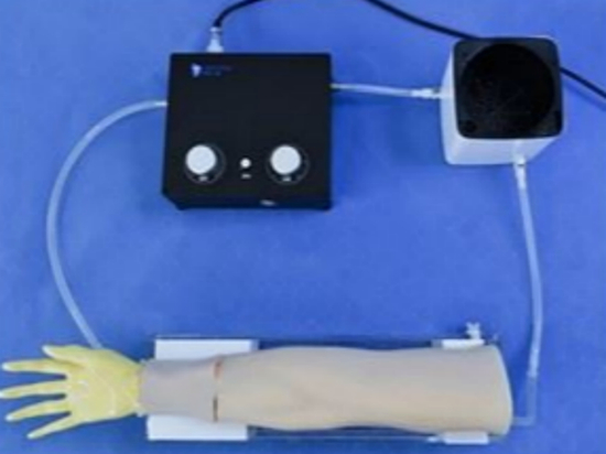
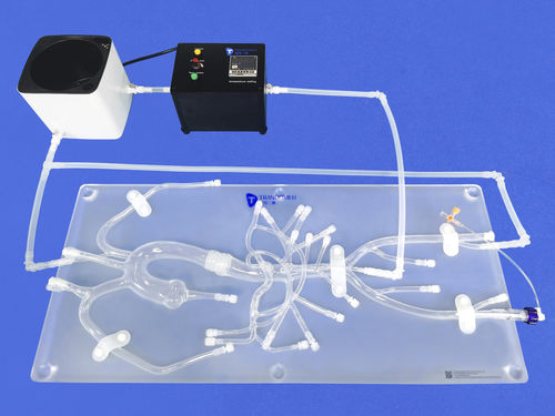
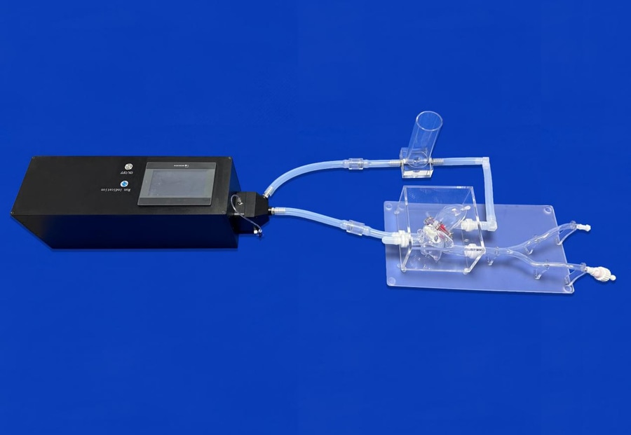
The Trando 3D printed valve model, combined with a proprietary hemodynamic system, generates highly realistic, adjustable pressure waveforms, helping researchers measure transvalvular pressure gradient, effective orifice area (EOA), and regurgitation flow in real time.
This helps researchers obtain performance parameters close to those in the human body, providing reliable data for the development of artificial valves.
II. Helping to Reduce the "Hidden Costs" of Leaflet Design
Insufficient leaflet opening or asynchronous closing often only becomes apparent during lengthy experiments, resulting in higher hidden costs.
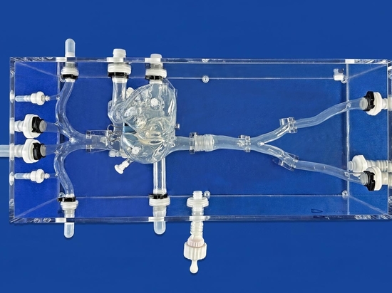
Trando 3D utilizes specially customized high-transparency models, combined with high-speed photography, to observe leaflet kinetics. This helps developers quickly identify stress concentration areas, optimize leaflet cutting schemes, and reduce development time and costs.
III. Solving the "Passability Challenges" of Delivery Systems
TAVR delivery devices struggle to accurately reproduce the resistance of the complex aortic arch in simulated pathways.
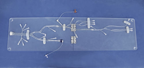
Trando 3D's customized aortic model is a 1:1 reconstruction based on real patient CT/angiography data, including realistic anatomical curvature. By linking to the pulsatile system, it can more realistically simulate the entire process of transvalvular, release, and retrieval, accurately assessing the delivery device's compliance and valve deployment performance.
IV. Helping to Detect Paravalvular Leaks (PVL) and Other Issues Early
Paraval leaks, a clinical concern, are difficult to reproduce on insufficiently simulated uniform valve annulus models. Trando 3D provides models with varying degrees of calcification to recreate irregular valve annulus morphology. The skirt's filling effect on calcified gaps can be tested in the laboratory stage, improving the leak-proof design.
V. Addressing the "Lack of Hands-on Experience" in Demonstrations and Training: Pure theory or 2D images make it difficult for doctors to intuitively experience the physical feedback of the device. Trando 3D customized heart valve model, 3D printed from angiographic data and combined with related hemodynamic systems, can highly simulate the human physiological environment, helping clinicians conduct accurate functional tests.
It is more suitable for advanced teaching and demonstrations, helping to shorten the learning curve for doctors and build trust in the product.