#Product Trends
3D Printed Lower Extremity Artery Vascular Simulation Model
3D Printed Lower Extremity Artery Vascular Simulation Model
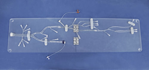
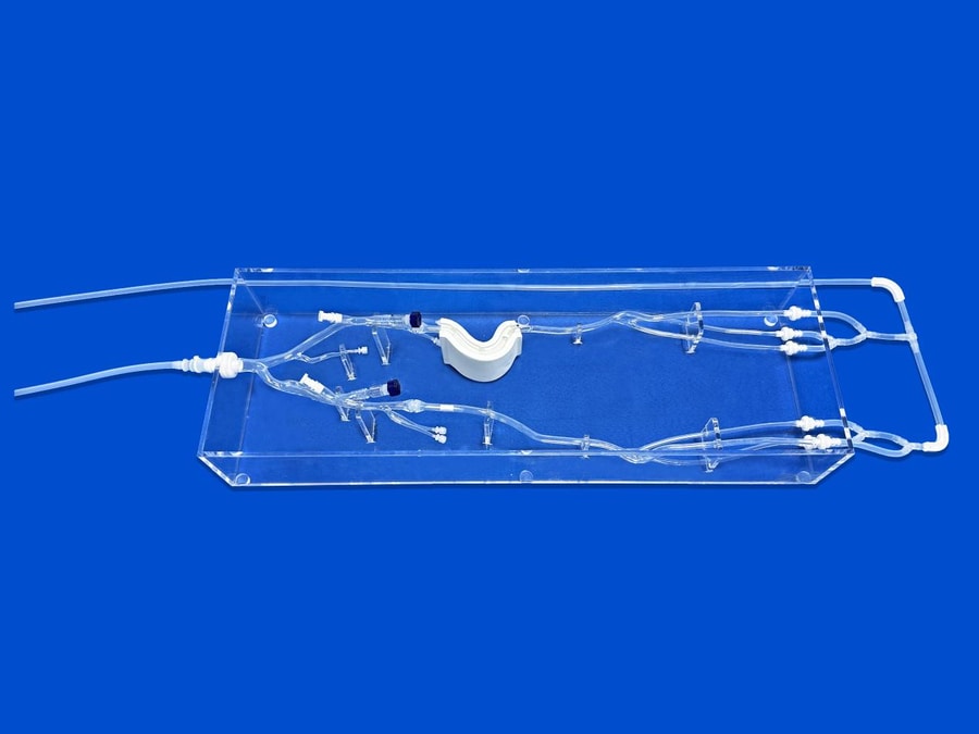
Vascular interventional training faces a long-standing bottleneck: rigid plastic phantoms oversimplify human anatomical variations, while animal models carry high costs, ethical restrictions and inconsistent vascular dimensions. Our newly upgraded 3D printed lower extremity artery simulation model completely solves this pain point by adopting authentic human CT scan data as the core reconstruction source. The whole vascular tree covers the abdominal aortic bifurcation, bilateral iliac arteries, superficial femoral arteries, popliteal arteries and distal tibial-peroneal branches, with a strict 1:1 scale restoration of natural lumen diameters, vessel tortuosity and branch distribution. The outer acrylic frame measures 908mm in height and 316mm in width, matching the real proportion of human peripheral arteries. Every vascular segment retains clinically critical dimensional parameters, ranging from the maximum aortic inner diameter of 16.0mm down to distal foot branches of 4.3–5.3mm, replicating the full spectrum of vessel calibers clinicians encounter daily. Unlike generic standardized models, this CT-based design eliminates artificial anatomical distortion, allowing trainees to feel identical spatial perceptions as real human angiography.
Complex peripheral artery disease (PAD) lesions are the core training focus of this phantom, with two interchangeable pathological modules embedded for targeted practice. First, replaceable calcification inserts mimic 65% stenotic plaques; each cylindrical calcification unit has an outer diameter of 8.0mm and inner residual lumen of 3.6mm, simulating hard atherosclerotic plaques that resist balloon dilation during angioplasty. Second, dedicated CTO (Chronic Total Occlusion) segments are integrated, with enhanced vessel tortuosity (maximum bend diameter 84mm) and integrated one-piece curved tubing without segmented connectors to restore the tight, twisted anatomical features of long occlusive lesions. Trainees can practice wire crossing, microcatheter support and balloon expansion under realistic lesion resistance, avoiding the over-smooth vessel surfaces of traditional simulators. The modular design enables rapid lesion swapping, so educators can assemble mild stenosis, severe calcification and full CTO scenarios within minutes for progressive training courses.
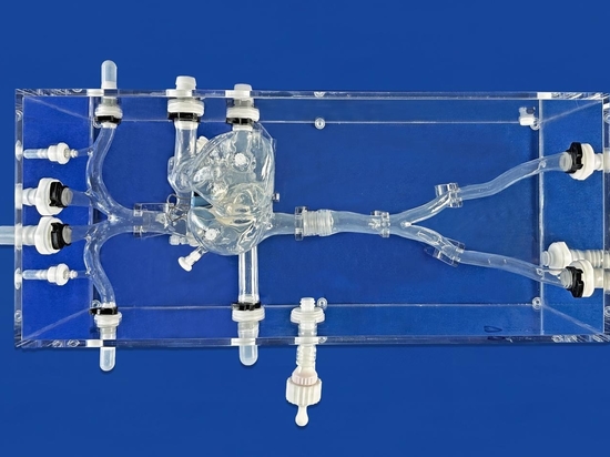
To replicate real interventional operation workflows, the model is equipped with four independent access ports compatible with 0–12F hemostatic valves, recreating common femoral artery puncture approaches for unilateral/bilateral simultaneous intervention. The entire vascular tree is fixed inside a transparent acrylic tank with customized support brackets, offering full visual observation of guidewire and catheter movement during procedures. A closed-loop perfusion circuit connects the phantom to a circulating pump: fluid flows downward from the top aortic inlet and circulates back to the pump via distal vessel outlets, simulating physiological blood flow pressure and flow velocity in lower limb arteries. The transparent shell lets instructors observe hidden wire looping, plaque contact and balloon expansion without fluoroscopy, significantly reducing radiation exposure during training. This complete fluid circulation system makes the phantom suitable for stent deployment, atherectomy and post-dilation skill drills.
This 3D printed lower extremity artery phantom serves three core clinical scenarios: standardized trainee training for vascular surgeons and interventional radiologists, pre-procedural complex case rehearsal, and medical device product demonstration. By reproducing real PAD anatomical and pathological features, it shortens the learning curve for junior physicians mastering CTO crossing and calcified lesion treatment. For medical manufacturers, the interchangeable lesion modules support bench testing of new guidewires, balloons and peripheral stents. Developed by Ningbo Trando 3D Medical Technology, the model balances anatomical authenticity, modular flexibility and long-term reusability, becoming a cost-effective alternative to animal and cadaver training resources.